Fossil fungi
Fossil fungi is a highly intricate subject,
investigated by only few palaeobotanists, although they represent an
important
component of ancient ecosystems [1,2]. Much work on fossil fungi has
been done lately but some of the
fungi and related phenomena reported here are not covered by the recent
monograph Fossil Fungi [4].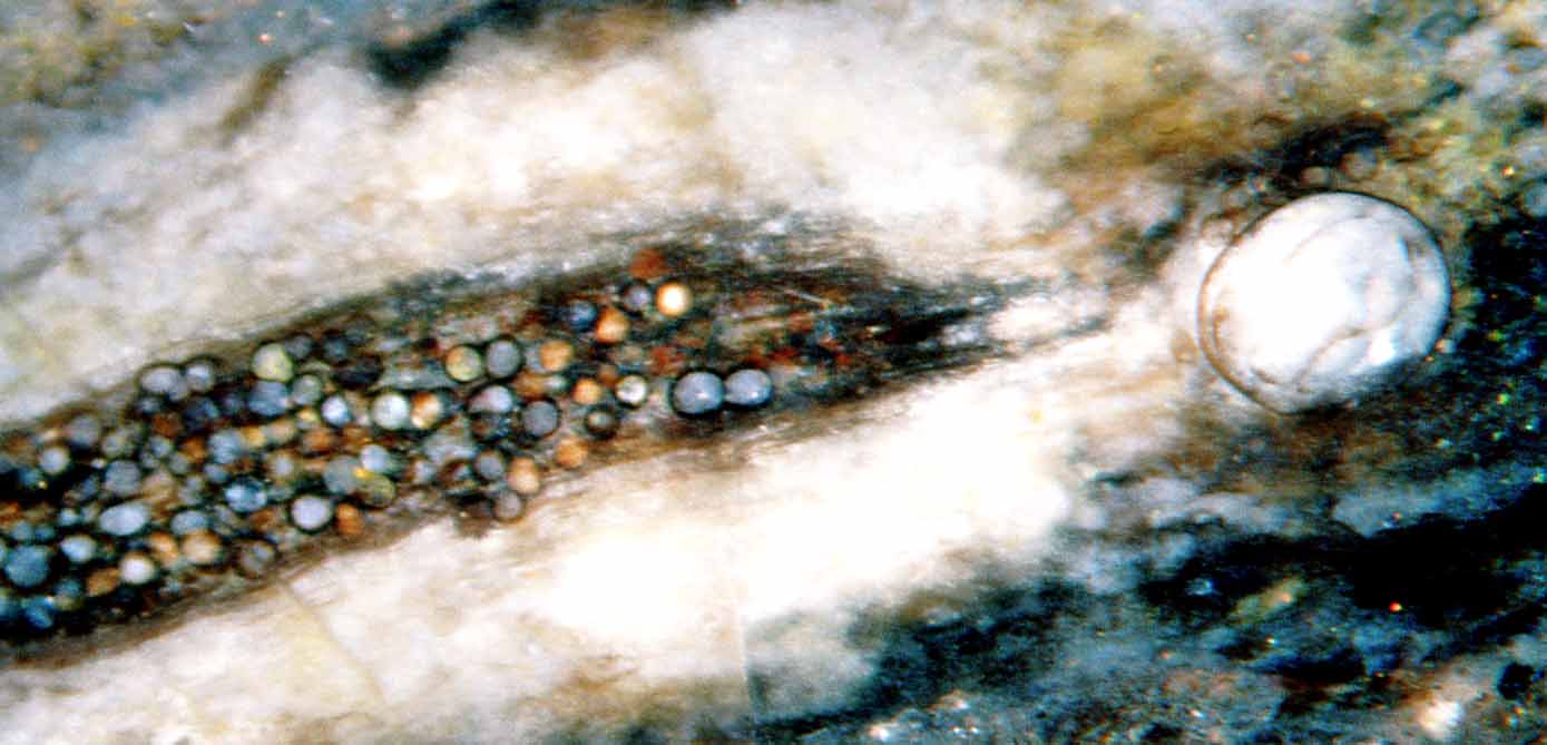
The delicate hyphae are seldom preserved, not even
in chert, with the
exception of the Lower Devonian
Rhynie chert, where they are abundant and represent
several species. More suitable for preservation are the spherical
or ovoid objects called resting spores, chlamydospores, or vesicles. A
diverse
collection of these is shown in a comprehensive early publication on
fossil fungi in the Rhynie chert [3]. Some are not
named but only numbered there, and
apparently some of them have not yet got a scientific name
hitherto. Hence it seems useful to look for more details related to
fungi in chert.
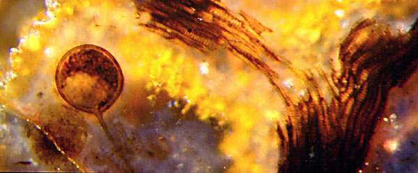
left: Fungus chlamydospore and hypha near forking xylem strand of
the early land plant Trichopherophyton
; transparent chalcedony with yellow precipitate and quartz
crystals in the background, Rhynie chert. Image width 1.4mm.
above: Fungus chlamydospores of two largely differing sizes
within plant debris in Rhynie chert. Image width 2mm.
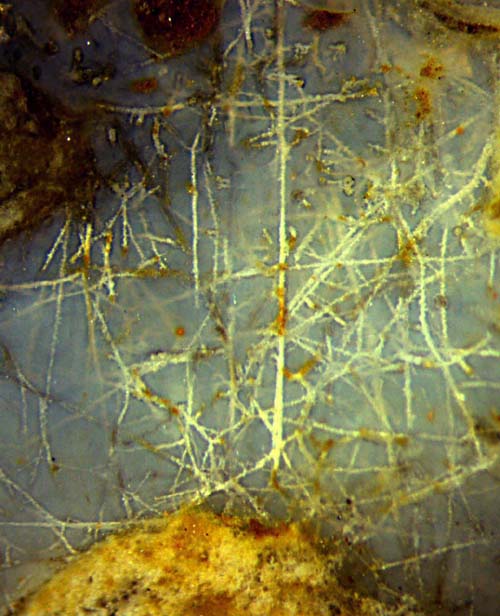
right: Fungus hyphae, interconnected, well
preserved in Rhynie chert.
Picture width 2.5mm.
The fungus-related phenomena described here are listed below
along with the number of the contribution in Rhynie
Chert News and Permian
Chert News.
Every number is linked to the related text.
Rhynie
Chert News
4
fungus-induced void pattern on cross-sections of Rhynie
chert plant
19
small chlamydospores within big one in decayed Rhynia
21
fungus-induced
void pattern on twin cross-sections of Rhynie chert plant
28
fungus-affected Asteroxylon
cross-section with dark cell fills
32
symbiotic fungus in Rhynie
chert plants forming dark clots in cells
54
additional evidence for fungus-induced void pattern formation
55
funny fossil fungus formations
63
several manifestations of Devonian fungi
76
symbiotic fungus in Trichopherophyton
forming dark clots in cells
77
stepwise silicification deduced from hypha
coating thickness
78
hyphae curved and surprisingly straight
84
miniature fossil sewage tank
85
cell-size dark clots often mistaken for mite coprolites
87
hyphae with microbial debris and thick coating
104
instructive examples of fungus resting spores
108
clusters of chytrid (?) zoosporangia resembling but differing
from Trewinomyces
109
wavy hyphae resembling the extant mycoparasite
Trichoderma
111
more wavy hyphae like Trichoderma
113 microbial formations
with cluster of Zwergimyces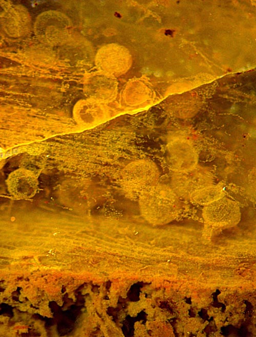
115
Zwergimyces
globules densely
clustered
116
fungus-related
spheres that come in pairs
117
fungus-induced voids on cross-sections
119 fungus-related
spheres with
fancy fills
120. fungus-related
spheres with alga connection
124. Asteroxylon
tissue affected by fungi
130.
Aglaophyton
hollow straw twin sections
142.
hyphae with thick silica gel coatings
143. branched hyphae with thick silica
gel coatings
145. tangles of hyphae with silica
gel coatings
148. more
wavy hyphae like Trichoderma
153.
nematophyte or fungus ?
160.
straight and wavy hyphae in Aglaophyton
163.
big chlamydospores with coloured fills
173.
surprisngly straight hyphae
165.
parasite scabs on Rhynie chert plants
177.
big glossy chlamydospores
186. conspicuous big
ellipsoid-shaped chlamydospore in hollow Aglaophyton
187. Symbiotic
fungus
Glomites in
the early land plant Aglaophyton
188.
Rhynia twice affected: first by parasite, then by Glomites
190. Fungus-related fossil spheres
in Rhynie chert
Permian
Chert News
14
hyphae with multiple coatings
17 microbes, hyphae,
chlamydospores, Scolecopteris
Picture on the right: Fungus chlamydospores in
decayed Permian plant matter. Image width 2mm.
Most of the cell-size dark clots in
Palaeozoic plant fossils thought
to be mite coprolites are no such but fungus formations instead.
Nevertheless the "mite coprolites" have got
widespread popularity among palaeobotanists who, by frequent mutual quotation, created a semblance of their reality. That problem has been separated here. It is
dealt with under Misconceptions, Oribatid mite coprolites,
and under "Wood
rot or coprolites".
H.-J. Weiss
2011, 2015, 2017, 2018, 2020, 2022, 2023
[1] T.N.
Taylor, J.M. Osborn: The importance of
fungi in shaping the paleoecosystem.
Rev. Palaeobot. Palyn.
90(1996), 249-262.
[2] D. Redecker:
New views on fungal evolution based on DNA markers and the fossil
record.
Res. Microbiology 153(2002),
125-130.
[3] R.
Kidston, W.H. Lang : On Old Red Sandstone
plants showing structure ... Part V. The Thallophyta ...
Trans. Roy. Soc. Edinburgh 52
(1921),
855-902.
[4]
T.N.Taylor,
M. Krings, E.L. Taylor: Fossil
Fungi. Elsevier 2015.