A dark side of palaeobotany
A dark side of palaeobotany has developed
nearly unawares during the latter two or three decades. It is centered
around
cell-size black clots in damaged or intact plant tissue, notably in
petrified wood. Contrary to common sense, the clots have been offered
as coprolites of never-seen mites or unknown creatures. This
inconsistency
did not bother those in the small circle of insiders who mutually
exchange and read their own publications.
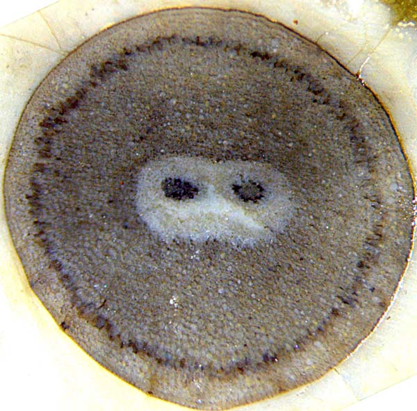
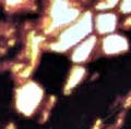
The mesmerising face shown here should serve as a warning to those who
still promote the idea of dark fills in cells being coprolites.
Fig.1:
Early land plant Aglaophyton,
seldom seen
well-preserved cross-section, width 4.4mm,
with forking central strand.
The cells with dark
fills, arranged as a ring, contain the symbiotic
fungus Glomites
rhyniensis [1].
Note also the cracks being deflected at the surface, indicating the
presence of a waxy cuticle typical of land plants.
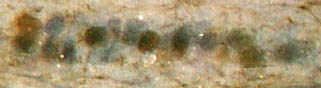
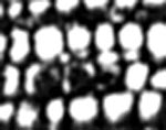
Fig.2: Aglaophyton
with dark
fills of cells as in Fig.1,
here radial lengthwise cut.
Figs.1,2: Rhynie chert, Lower Devonian.
It is unbelievably absurd to assume
that cell-size clots resembling those in Figs.1,2 are
coprolites. Fungus hyphae are known
to be able to enter into cells and form a clot there by
prolific branching, known as an arbuscule [2].
In order to demonstrate the absurdity of the widespread coprolite
hypothesis, a few details have been taken from the scientific
literature. The authors
[3-7] and several others do not wonder how the
alleged fecal clots got into cells, occasionally one beside another
in a row (Figs.3,6,11,12).
Clots which fill the angular cell cross-section are
shaped
accordingly but smaller ones can be nearly
spherical. Apparently they
are stiffer than the degraded tissue so that they retain their shape
when
the empty cells are squeezed (Fig.4).
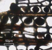
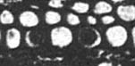
Fig.3:
Rows of globular black clots in Permian wood from China, not filling
the cell
cross-section but apparently being attached to the wall: alleged
coprolites in [3], Fig.4F.
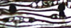
Fig.4: Longitudinal cut of Permian wood, slightly squeezed tracheids
with stiff clots inside: alleged coprolites in [3], Fig.4D. (See
also Fossil
Wood News 5,
8.)
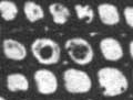
 Figs.5-9:
Clots in Permian wood from Thuringia, Germany:
Figs.5-9:
Clots in Permian wood from Thuringia, Germany:
Fig.5 (far left): Psaronius
phloem cells completely filled
with dark substance: alleged coprolites in [7];
Figs.6,7: alleged coprolites in [4] Abb15: row
of clots (Fig.6), clot with pentagonal contour
(Fig.7), similar cells
and one big flat-sided clot nearby;
Figs.8,9: "Twin clots": alleged coprolites in
[5],
Fig.1C.
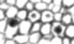
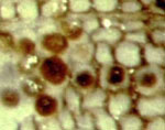
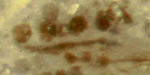
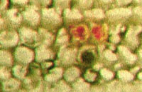
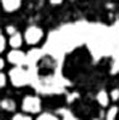
Fig.10-13: Clots in Permian wood from Germany:
Fig.10: Wetterau, Hesse, alleged coprolites in [6];
Fig.11,12: Schallodenbach, Rhine-Palatinate, own sample.
Fig.13: Döhlen basin, Saxony, own sample.
The professionals fond of the elusive mites [3-7] would
doubtless misinterpret the few own samples
(Figs.11-13) which essentially do not
differ from theirs in
Figs.3-10. Fig.12 is exceptional since the clots are still in their
original position although the cell walls have disappeared. In
Fig.13, hyphae seem to have grown between the cells before one of
them supposedly entered into a cell and branched profusely into a clot
there,
which eventually turned dark.
It is not known whether there is a connection to the hematite
above.
Although it is
nearly self-evident that clots inside plant cells cannot be coprolites, additional evidence from the Rhynie chert (Figs.1,2) may
help to convince those who would not see the simple truth.
Own samples: Fig.1: Rh2/73.1, obtained from J. Shanks,
Rhynie 2002, Fig.2: Rh2/83.2
Fig13: Bu7/111.2
Figs.11,12:
Sch/3.1, obtained from Ch. Krüger,
Schallodenbach, Rhine-Palatinate, Germany
H.-J.
Weiss
2015
[1] T.N. Taylor
et al.: Fossil arbuscular mycorrhizae from the Early
Devonian,
Mycologia
87(1995), 560-73.
[2] T.N.
Taylor, E.L. Taylor, M. Krings : Paleobotany, Elsevier
2009, Fig.3.96.
[3] Zhuo
Feng, Jun Wang, Lu-Yun Liu :
First report of
oribatid mite
(arthropod) borings and coprolites in Permian woods from ... China.
Palaeogeography,
Palaeoclimatology, Palaeoecology 288(2010), 54-61.
[4] R.
Rößler, R. Kretzschmar, Z. Feng, R. Noll:
Fraßgalerien von
Mikroarthropoden in Konifernhölzern des frühen Perms von Crock,
Thüringen.
Veröff. Mus.
Naturkunde Chemnitz 37(2014), 55-66.
[5] Zhuo
Feng,
J.W. Schneider, C.C.
Labandeira, R. Kretzschmar, R.
Rößler:
A specialized feeding habit
of Early
Permian oribatid mites.
Palaeogeography,
Palaeoclimatology, Palaeoecology 417(2015), 121-124.
[6] K.
Goth, V. Wilde : Fraßspuren in permischen
Hölzern aus der Wetterau,
Senckenbergiana letaea
72(1992), 1-6.
[7] M.
Barthel, M. Krings, R. Rößler: Die schwarzen Psaronien von
Manebach, ihre Epiphyten, Parasiten und Pilze.
Semana 25(2010), 41-60.
 |
 |
85 |