Fungi in Permian chert
For variuos reasons, fossil fungi
are rare despite of the fact that they had been helpful to plants when
they occupied the dry land in the Silurian and Devonian [1].
As an exception, the Rhynie Chert (Lower Devonian) is famous among
biologists for the excellent preservation of hyphae and
"chlamydospores" of several fungi.
Only
a few of the expected numerous palaeozoic fungus species have been
found well preserved in chert, and only part of them have been
scientifically described. Some lived as symbionts or parasites, others
lived on decaying dead plant tissue, also in the swamp water where they
occasionally formed extended tangles of branching hyphae. With
silica-rich swamp water turning into gel and finally into chert, the
hyphae became silicified.
Cherts from Germany
have yielded very few fungi. It can be expected that some of the
numerous chert samples recovered from Döhlen basin (Lower
Permian), superficially or not yet
inspected hitherto, will provide a few more specimens. Hyphae within
quartz crystals (Tertiary) have been found near Warstein [2].
A
peculiar fact may be mentioned here: A particular kind of wood rot with
hyphae filling the wood cells with profusely branched "arbuscules" has
been described by several palaeobotanists, with increasing zeal, as
oribatid mite coprolites in petrified wood.
(See Rot
or "coprolites" and Research gone astray.)
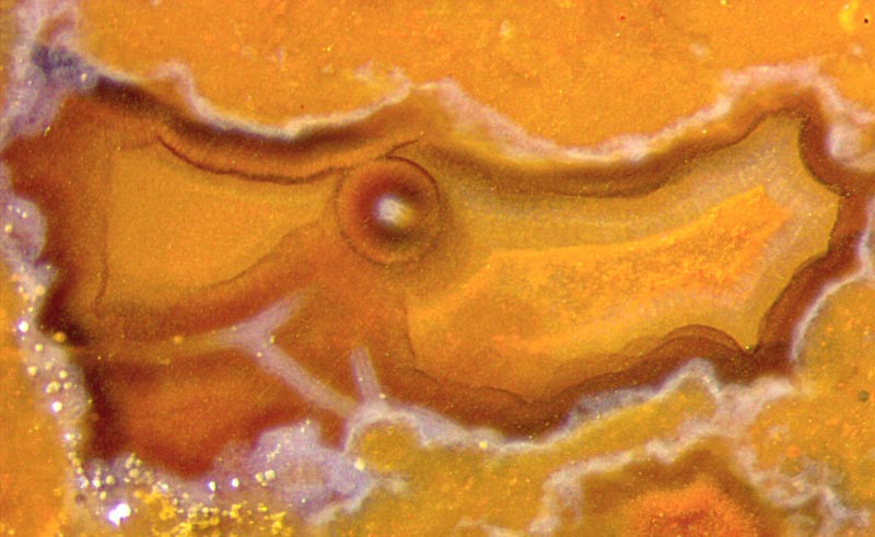 Fig.1:
Chalzedony, formerly water-filled cavity with poorly visible fungus
hyphae, diameter <10µm, with pale coating, about 25µm, and brown
coating, 170µm. Picture width 1.4mm.
Fig.1:
Chalzedony, formerly water-filled cavity with poorly visible fungus
hyphae, diameter <10µm, with pale coating, about 25µm, and brown
coating, 170µm. Picture width 1.4mm.
The lower pH inside decaying plant parts in
silica-rich water favours silica gel formation
there first. Subsequently, coatings may be formed.
Fungus hyphae often thrive in the remaining water-filled cavities. They
become coated with silica gel, eventually with several layers due to
changing parameters, as temperature. The concentric layers may appear
as pale in clear vicinity or clear in pale vicinity, for example
(Fig.1-3). The coated hyphae may bend down under their own weight.
The subsequent processes
are governed by the supersaturation of the remaining solution: High
supersaturation makes gel and later chalzedony in the whole cavity. Low
supersaturation makes quartz crystals on the coated hyphae and on the
walls. Crystal growth ends when silica does not
enter by
diffusion (not flow !) anymore. Later the water vanishes and the coated
hyphae cross the empty cavity. (See
Rhynie
Chert News 63,
64,)
Coated
needle-shaped crystals and mineral formations like those in moss agate
are liable to being confused with hyphae. Long straight parts ending at
branching sites are absent in moss agates, and curved parts are absent
with crystal needles (Fig.1).
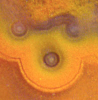
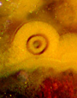
Fig.2,3: Multiply coated fungus hyphae, height of the pictures 0.35mm,
same scale as Fig.1.
Diameters of the coatings in Fig.2 (µm):
10, 14, 22, 28, 40, 48, 110, 125,
145(?).
Fig.4
(below): Tree fern frond stalk, largely decayed
and silicified while lying in swamp water, indistinct tangle of poorly
preserved fungus hyphae in some places, 4 (or more)
spherical chlamydospores, cut or transparent, empty, thick
microbial cover (cyanobacteria ?) on the left. Width of the picture
5.5mm.
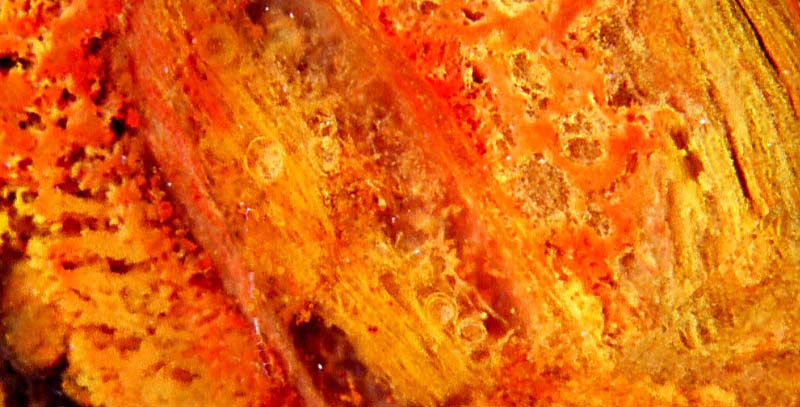
Fungus hyphae are rare in the cherts of the Döhlen basin.
Probably
they decayed before they became coated with silica gel. The
comparatively
big spherical chlamydospores, which possibly guarantee
survival after decay of the hyphae, are another feature revealing the
presence of fungi. They are not easily seen among the debris in Fig.4.
Here, the wall of the chlamydospores
is apparently not much stained by the abundant yellow or red iron
oxides deposited from soluble iron compounds.
(In Permian cherts, chlamydospores seem to be much less
abundant and less clearly seen than in the Lower Devonian cherts from
Rhynie.)
Samples: Döhlen basin, Lower Permian.
Fig.1-3: Kleinnaundorf,
Kohlenstr., found by
H. Ahlheim;
cut and polished by H. Albrecht, label Bu7/207.
Fig.4: own find, Hänichen, Käferberg; own
collection, label H/333.1 .
H.-J.
Weiss
2015 modified 2017
[1] D. Redecker: New views on fungal evolution based on
DNA markers and the fossil record.
Res. Microbiology 153(2002), 125-130.
[2] M. Kretzschmar: Fossile Pilze in Eisen-Stromatolithen von Warstein.
Facies 7(1982), 237-259.
|
 |
 14 14 |