Funny fossil fungus formations
The Lower Devonian Rhynie chert is known not only for
the well preserved early land plants but also for the abundance of
fungi preserved therein [1]. By presenting a
few examples selected from a larger number, this
contribution is meant to draw
attention to the variety of phenomena brought about by fungus activity.
Some phenomena are caused by a fungus species well known and
described in the palaeobotany literature [1,2]. An example is seen in
Fig.1 where cells with dark fills
representing so-called arbuscules of the fungus
Glomites rhyniensis
are seen loosely arranged in two nearly circular
lines.
The formation of arbuscules by several
recent species of Glomus
is
a widespread phenomenon known as arbuscular mycorrhiza, which is
important in modern plant life and agriculture.
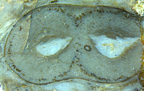
Fig.1: "Burglars' mask", cross-section of forking Aglaophyton at a
position between the separation sites of xylem strand and shoot, with
several features indicating fungus activity.
Obviously,
the cut plane shown in Fig.1 is positioned above the forking site of
the central
strand but below the forking site of the shoot
so that two separate strands are seen but no separate prongs of the
shoot. The cells with dark fill are a common sight on
circular cross-sections, where they form a circle keeping at a small
distance from the epidermis. Here they are tending to form two circles
as if anticipating the impending
separation of the shoot into prongs. This raises the question what
governs the position of the fungus-affected cells. Obviously their
position is determined by a more complex rule than simply by an equal
distance from the epidermis as one could suspect from the usual aspect
of circular sections. Hence, there is a problem whose solution might
contribute to a better understanding of this particular type of
symbiosis.
The most conspicuous feature in Fig.1, the
big voids immediately beneath the (deformed) central strands, are
interpreted here
as a growth anomaly caused by a fungus. There is no straightforward
evidence for this but comparison with more uncommon voids and related
argumentation may be convincing. (See Rhynie
Chert News 4,
21,
54.)
Voids formed by mere decay would most probably not be positioned
symmetrically. The observations strongly suggest that one or more
fungus species had affected the live plant. Another
feature indicating the presence of a fungus is a
chlamydospore, which is a common but not yet thoroughly understood
fungus formation, seen on the right half. More chlamydospores are seen in
Figs.2-5 below. They are similar to but differ
from Palaeomyces
Gordoni [3].
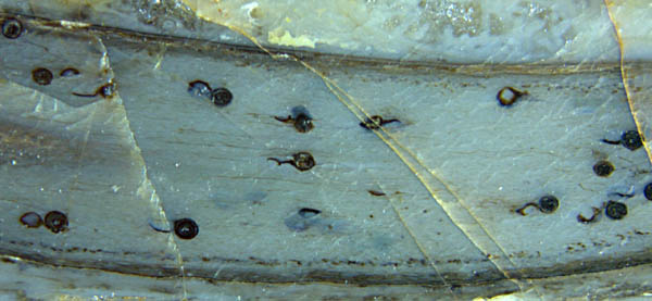
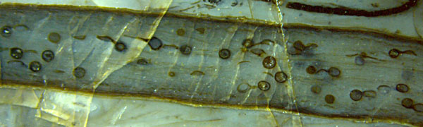
Figs.2,3,4: "Tadpole
race" in Aglaophyton:
Fungus chlamydospores of uncommon aspect due to attached "tails" which
are the remains of empty vesicles. Note also the line of small dots
marking the presence of Glomites
and a few very thin hyphae aligned lengthwise in Figs.2,4 below.
While the fungus "body" consists of a dainty system
of hyphae hardly visible here,
the chlamydospores
or resting spores seen here look like strong-walled spheres, often with
the remains of same-size objects attached
(Figs.4,5). The latter feature is less common
in the Rhynie chert.
Usually the
chlamydospores are attached to a thin
hypha which is seldom seen on arbitrarily positioned cut faces. (See here).
In
this sample the chlamydospores had been or are
still attached to an enigmatic vesicle, apparently by a short thick
tube (Fig.5),
seen in various stages of decay (Figs.2-4). (See
Addendum below, also Rhynie
Chert News 64
Fig.9.) Such feature is not clearly
seen in the detailed early work on Rhynie
chert fungi [3]. 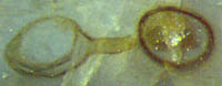
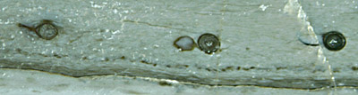
Fig.5: "Spectacle",
fungus chlamydospore connected to an enigmatic vesicle.
Detail of Fig.3, width 1mm.
Fungus hyphae like those faintly seen as very thin dark
lines in Figs.3,4 are
usually quite abundant in the Rhynie chert but rare in other cherts. If
coated with one or more layers of chalcedony or quartz they may appear
as conspicuous networks, even as free-standing ones in cavities. In
Fig.6 the hypha is hardly seen as a tiny brown
dot in the "eye",
nevertheless it has turned the random assembly of cracks and
whithish precipitate into a kind of chimera.
As a funny coincidence, the tiny hypha coated with clear chalcedony
appearing dark under incident light had aroused the curiosity leading
to closer inspection which drew the attention to some dots in front of
the chimera, which resulted in the discovery of the oldest
rotifer ever seen. (See Rhynie
Chert News 23.)
Hence one can say that without the coated hypha the
Devonian rotifer would probably not have been found up to now and would
not be found for
some time to come.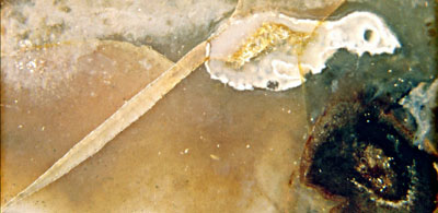
Fig.6: "Chimera" in Rhynie chert, composed of cracks, whitish
precipitate, and a fungus hypha with
thick coating of clear chalcedony appearing as a dark eye. Image width 7mm.
Finally
it can be stated that the Rhynie chert is exceptional also for its
preservation of fungi, which justifies the expectation that more
details will be found and put together into a consistent reconstruction
of the complex web of early plant and fungus life involving symbiosis
and degradation of organic matter.
Samples: Figs.1-5: Rh6/17, 2002 gefunden. Fig.6: Rh12/83.2 (90g), found by S. Weiss in 2006.
H.-J. Weiss
2013, emended 2014
Addendum 2014
What
is called chlamydospores in Figs.2-5 has been thoroughly described in
[4] as "acaulosporoid glomeromycotan spores" forming "spore-saccule
complexes". The spore is thought to develop sideways on the tail of a
saccule. This means that the visual impressions from Fig.5 above and
Fig.9 in Rhynie
Chert News 64
, which suggest a straight connection by a thick tube, should
be
regarded as an illusion due to a particular line of vision. Apparently
the deeper question why a big spore develops from an equally big
saccule on a thin hypha is dealt with in the ample fungus literature
referred to in [4].
[1] T.N.
Taylor, J.M. Osborn: The imporance of
fungi in shaping the paleoecosystem.
Rev. Palaeobot. Palyn.
90(1996), 249-262.
[2] T.N. Taylor,
E.L.Taylor, M. Krings: Paleobotany, Elsevier 2009.
[3] R.
Kidston, W.H. Lang : On Old Red Sandstone
plants showing structure ... Part V. The Thallophyta ...
Trans. Roy. Soc. Edinburgh 52
(1921),
855-902.
[4] N.
Dotzler, Ch. Walker, M. Krings, H. Hass, H. Kerp, T.N.
Taylor, R. Agerer:
Acaulosporoid
glomeromycotan spores with a germination shield from ... Rhynie chert.
Mycol. Progress (2009) 8,
9-18.
 |
 |
55 |