Parasite scabs on Rhynie chert plants
The Early Devonian habitat preserved in the
Rhynie chert is well known among biologists not only for its early land
plants but for its abundance of fungus species as well [1]. A very
common
symbiotic fungus in the early land plants
appears as dark fills of cells in a concentric ring on cross-sections,
here faintly seen in
Fig.1. The majority of fungi in the Rhynie chert
are saprophytes. Their hyphae and resting spores seen inside and
outside decaying plants are quite common. Quite uncommon is the
organism seen here, which apparently causes live plants to grow black
mounds without doing much damage.
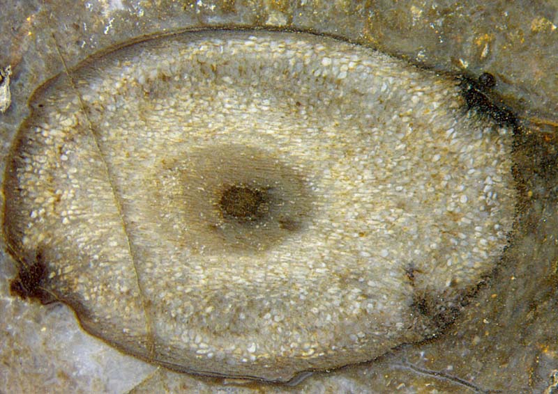
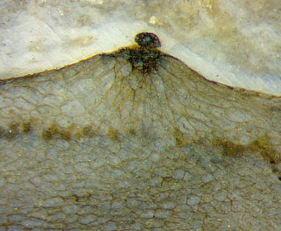
 Figs.1,2: Tilted cross-section of well preserved Aglaophyton, invaded
by an unknown organism interfering with cell growth and
producing dark matter.
Figs.1,2: Tilted cross-section of well preserved Aglaophyton, invaded
by an unknown organism interfering with cell growth and
producing dark matter.
Frame widths 7mm and 2mm.
As a characteristic feature of the invaded spot, the dark misshapen
tissue produces a globular body on top as if it were a propagule. A
similar spot is seen on the
left of the section in Fig.1. Possibly a globular body had been shed
there earlier.
The control of cell growth of Aglaophyton by the
parasite is most obvious
in Fig.3. Cells in a near-surface region had been induced to grow such
that they form a regular mound. The globular objects on top seem to be partially translucent,
particulary the one in Fig.5.
It is
not known in which way the cavity below the dark spot in Fig.4 may be
related to it.
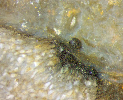
The symbiotic fungus
Glomites
rhyniensis [1] seen as
brown fills in Fig.3 and the parasite on the mound apparently do not
mutually interfere.
Annotation: Dark fungus matter inside cells had given rise to a
veritable craze of its
interpretation as mite coprolites: See Rhynie
Chert News 85 .
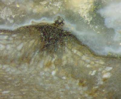
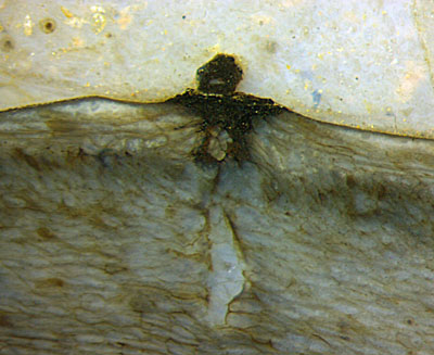
Figs.3-5: Mounds grown on the surface
of Aglaophyton due
to a parasite interfering with cell growth.
Frame widths 2mm.
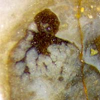
Fig.6: Parasite with globule on the surface of Rhynia. Frame
width 1mm, same scale as Figs.2-5.
The tissue in Fig.6 is poorly preserved so
that an infection of live Rhynia
is not evident here. Doubtless it is Rhynia
because there are no other plants in
this sample. Globules like these must not be confused with the often
abundant warts on Rhynia (Rhynie
Chert News 159),
which may appear as nearly globular in rare cases.
In view of other cases of
plant growth being interfered with by fungi, this parasite, too, is
probably some fungus.
Samples: Figs.1,2,5: Rh2/237.2 (0.2kg)
found in 2014; Fig.3:
Rh2/4.3 (2kg) obtained from Shanks
in 1998; Fig.4: Rh2/73.2 (0.29kg)
obtained from Shanks
in 2002; Fig.6:
Rh22/4.2 (70g) found at Castlehill in 2009.
H.-J.
Weiss 2020
[1]
T.N. Taylor, M. Krings, E.L. Taylor: Fossil
Fungi. Elsevier 2015.
 |
 |
165 |