Nematophyte or fungus ?
Now that the famous nematophyte Prototaxites is
interpreted as a huge fungus or lichen [1] and the less known small Nematophyton taiti
[2] from Rhynie has been re-named Prototaxites taiti
[3], the startling question arises whether or not more nematophytes are
really fungi or lichens. This question will certainly be answered some
day but for the present we will put it aside and delve into details
related to it.
So
it may be appropriate to look at
the own specimen of a fossil nematophyte
resembling Nematophyton
taiti [2], tentatively described in Rhynie
Chert News 46
(and [4]), once more. It
is readily assorted with the nematophytes by the tangle of curved tubes
(Fig.1) in the middle layer of the sandwich structure seen in
Rhynie
Chert News 35.
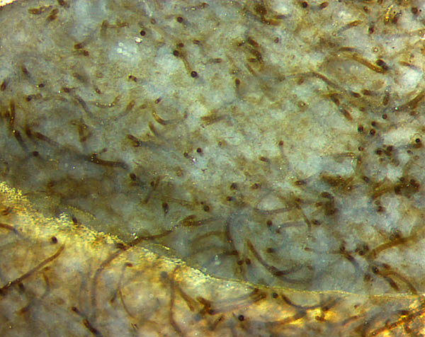
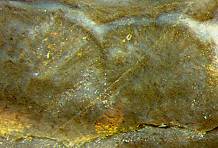
Fig.1: Tangle of tubes with various diameter and curvature
in the
middle layer of a flat nematophyte. Width 2mm.
As seen here, the tubes do not seem to
follow any rule, not even randomness. Their diameters vary between 10µm
and 25µm but most of them seem to be in the
range of 17-20µm.
From the absence of mineral debris one may conclude
that the tubes did
produce organic gel as it is known from other nematophytes, with the
purpose of keeping the tubes together in a lump of gel and to
prevent exsiccation in case of dried-up surroundings.
It is not seen where the tubes come from. There are no conspicuous "branch-knots"
thought to be related
to tube generation, as known from Nematoplexus.
One may wonder whether the tubes are possibly generated
in the clusters on the right in Fig.1.
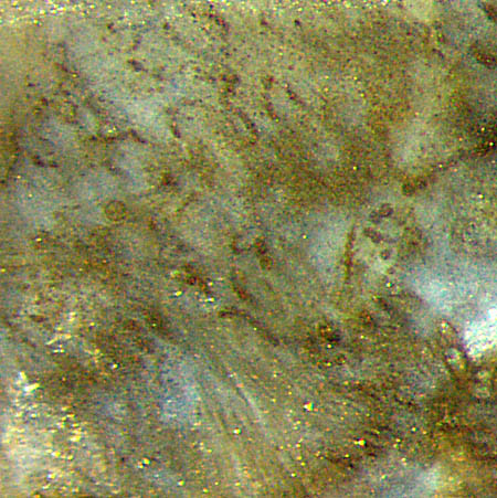
 Tube aggregates of a different type are rather
puzzling than
elucidating for several reasons. They appear as dark bundles of
thick-walled tubes, mostly seen as cross-sections in Fig.2,
which leads to the unexpected conclusion that
this nematophyte specimen, or the part of it seen here, must have a
preferred direction which is not apparent from the tangle of tubes in
the inner layer (Fig.1). (Also it appears that the chert sample had
been
cut into slabs incidentally such that most of the mentioned tube
bundles are seen as cross-sections.)
Tube aggregates of a different type are rather
puzzling than
elucidating for several reasons. They appear as dark bundles of
thick-walled tubes, mostly seen as cross-sections in Fig.2,
which leads to the unexpected conclusion that
this nematophyte specimen, or the part of it seen here, must have a
preferred direction which is not apparent from the tangle of tubes in
the inner layer (Fig.1). (Also it appears that the chert sample had
been
cut into slabs incidentally such that most of the mentioned tube
bundles are seen as cross-sections.)
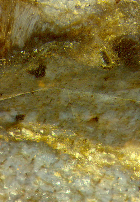
Fig.2 (left): Partial section of a flat nematophyte: tangle
of tubes in bluish gel below, former gap with mineral debris,
intermediate
layer with very narrow tubes and a few bundles of big dark tubes, outer
layer with
pockets of pale aligned tubes above left. Image
width 1.5mm.
There
is no indication where the dark bundles come from or what they mean.
They
are found scattered in the middle layer of the
nematophyte and also in
the outer layer with very narrow tubes, as seen
in Fig.2. The
number of tubes per bundle varies between 1 (near the middle of Fig.2)
and several dozens (on the right in Fig.2). The tube diameters in the
bundles, 7-27µm,
are similar to those in the middle layer (Fig.1).
Apparently the specimen had been torn before silicification
and muddy water with mineral platelets
had filled the gap and became silicified along with the organic gel
above and below.
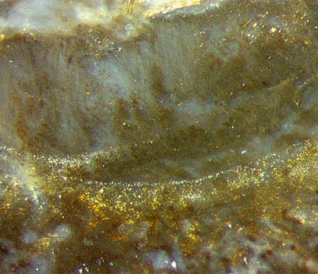
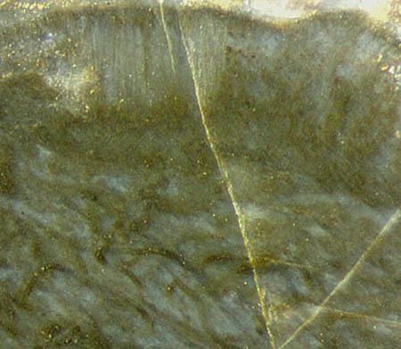
Figs.3,4
(right): Nematophyte, tangle of curved tubes below and pockets of
straight aligned tubes at the above surface, unidentified
structures in between, intermediate layer
smaller than in Fig.2. Width 1.5mm.
Fig.5 (below): Nematophyte,
straight narrow tubes arranged in a fan-like
way.
Figs.1-5: same
scale. Figs.2-5: width
1.5mm.
The straight narrow tubes usually seen aligned
in pockets along the surface are occasionally
arranged in a more or less divergent way, as shown
in Fig.5
and Rhynie
Chert News 46.
Unlike the curved tubes in Figs.1-4, the straight tubes in the pockets (Figs.2-5)
are
distinguished by
their definite lengths of about 0.5mm, less distinct small
diameters, and by their densely packed
arrangement with all tube ends together forming a common face
integrated into the nematophyte surface.
Strings
of beads, probably spores, are seen
among the straight tubes in the pockets, apparently
with one string per tube, thus differing from the specimen in [3],
where the spores are produced within wider asci by the hundreds.
Fig.6
(right): Spores (?)
of various sizes arranged in (mostly deformed) strings, bunch of empty
(?) aligned tubes below.
Image size 0.75mm, magnification twice that of
Figs.1-5.
Confusing as it may be, Fig.6 seems to offer the
interpretation that strings
of tiny spores had formed in the straight tubes directed towards
the surface, then expanded and became deranged while the tubes decayed.
There is still a bunch of orderly aligned tubes, directed
downward and apparently without visible dots inside, vaguely seen in
Fig.6 below.
As
a less probable alternative interpretation, the dark clots might be no
spores but alga cells which lived in a symbiosis with the
nematophyte as a lichen [5].
The dispute on "nematophyte or fungus (or lichen)" concerning Prototaxites is
analysed thoroughly and comprehensibly in [6]. An image of Nematophyton
taiti in
[7] is unsuitable for comparison with this
specimen and with [3] because of a scale error
(factor 2.4) and the mirror image.
Besides some differences between this specimen and the one
called Nematophyton taiti
[2] once but Prototaxites taiti [3] now,
their
structure is so similar that this one may be regarded
as an ascomycete fungus
with "nematophyte aspect" as well.
Sample: Rh2/7 (2.1kg), obtained from Shanks in 2000.
H.-J.
Weiss
2020
[1] F.M. Hueber:
Rotted wood-alga-fungus: the history and life of Prototaxites Dawson
1859. Rev. Palaeobot. Palynol. 116 (2001), 123–158.
[2] R.
Kidston, W.H. Lang : On Old Red Sandstone
plants showing structure ...,
Part V, Trans. Roy. Soc. Edinburgh 52
(1921),
855-902.
[3] R.
Honegger, D. Edwards, L. Axe, Ch. Strullu-Derrien:
Fertile Prototaxites
taiti: a basal ascomycete with
inoperculate, polysporous asci lacking croziers.
Phil.Trans. Roy. Soc. B 373 (2017): 20170146.
[4] H.-J.
Weiss:
Enigmatic Organisms - Insights derived from new
finds, Poster presentation, EPPC Budapest 2010.
[5] G.J. Retallack, Ed Landing:
Affinities and architecture of Devonian trunks of Prototaxites loganii.
Mycologia, 106(6) (2014), 1143–1158.
[6] H. Steur: Prototaxites.
Google: steurh.home.xs4all.nl/engprot/
[7] www.abdn.ac.uk/rhynie
 |
 |
153 |