Microbes, fungi, maggot fern
 Fungi
are supposed to have been around since the Proterozoic [1] but have
been preserved as fossils only under conditions of rapid
silicification, as in the Lower Devonian habitat turned into the Rhynie
chert
with lots of well-preserved fungus remains. Fossil fungi are rare in
the Permian cherts of the Döhlen basin.
(See Permian
Chert News 14.)
Perhaps some hyphae are easily
overlooked if poorly preserved, or they are mistaken for microbial
filaments or plant hairs.
Fungi
are supposed to have been around since the Proterozoic [1] but have
been preserved as fossils only under conditions of rapid
silicification, as in the Lower Devonian habitat turned into the Rhynie
chert
with lots of well-preserved fungus remains. Fossil fungi are rare in
the Permian cherts of the Döhlen basin.
(See Permian
Chert News 14.)
Perhaps some hyphae are easily
overlooked if poorly preserved, or they are mistaken for microbial
filaments or plant hairs.
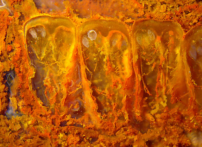
Fig.1: Curved pinnules of the "maggot fern" (Scolecopteris)
in a row, cut such that the cross-sections with sporangia and the
sheltered spaces with thin hyphae and thicker hairs are seen. Width of
the image 8mm.
A rare combination of microbes, fungus hyphae, and plant hairs is seen
in
Fig.1, which shows an inclined cut of a row of curved maggot fern
pinnules. Apparently these pinnules are even more curved than those
seen in side view in Permian
Chert News 1.
Owing to the
curvature, the pinnules appear nearly as
cross-sections above (see Permian
Chert News 15)
and provide a look into the sheltered space between the fringes of the
lamina below.
The pale empty sporangia of the fern may be easily recognized as such.
The thin fungus hyphae are easily seen against the dark background if
not
covered with microbes too much. The hairs sparsely distributed at the
lower side of the pinnules are much thicker than the hyphae (Fig.2).
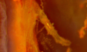
Fig.2:
Detail from Fig.1, near image centre, hair emerging from the lamina of
the pinnule, with thin fluffy microbial cover, glistening spot where
the
cover probably had been scratched off before silicification so that the
smooth reflecting surface appeared. Image width 0.7mm, lamina thickness
0.25mm.
The microbes involved here do not form filaments by
themselves but coatings and
floccules without definite shape but with sharp boundaries against the
surrounding water.
The
yellow, red, and brown colours are provided, as usual, by iron oxides.
The bluish hue on the left of Fig.1 is not due to a mineral pigment but
to light scattered on tiny precipitates in the clear chalzedony.
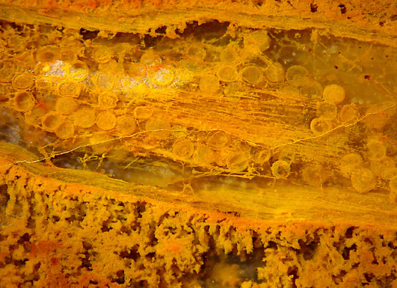
Fig.3:
Slightly inclined lengthwise section of a largely decayed root
surrounded by a thick fluffy microbial coating and with numerous
spherical chlamydospores of some fungus with a few thin hyphae standing
out
against the dark background. Width of the image 5mm.
Like
hyphae, chlamydospores are seldom seen in Permian cherts but are common
fossils in the Rhynie chert. (See Rhynie
Chert News 104, 115.)
The large bunch of parallel filaments is the decayed central strand
The assemblage of numerous chlamydospores
in Fig.3 is the only find of this type in the Permian
cherts of the Döhlen basin but similar
assemblages are known from the Rhynie chert. Diameters
of the spheres in Fig.3 are up to 0.3mm, thus being comparable to those
in 104
.
No attempt has been made here to assign
these finds to a certain
fungus clade.
Another problem remains: Why did this fungus, which is a tangle of thin
hyphae,
choose to produce bulky chlamydospores in large numbers and to
place them clustered together ? One may assume that there must be a
reason which has to be found out.
It may be appropriate here to
warn against a potential misinterpretation: Spherulites must not be
mistaken for chlamydospores. Two of such, size 0.34mm, are half seen in
Fig.1 between the first and second pinnule.
Samples: Döhlen basin, Hänichen, Käferberg.
Figs.1,2: H/321.1, 0.85kg, 1999,
Fig.3: H/333.2, 4.65kg, 2001.
[1] T.N.Taylor, M.
Krings, E.L. Taylor: Fossil Fungi. Elsevier 2015, p.28.
H.-J.
Weiss
2017
|
 |
 17 17 |