Fungus from Rhynie chert helps to refute Permian
/ Triassic mites hypothesis
As a common sight on cross-sections of Aglaophyton
(former
Rhynia major),
the most common plant in the Rhynie chert, there are cells
with dark
content, loosely arranged as a concentric ring. The phenomenon has
been described in detail and explained as being due to the fungus
Glomites rhyniensis
[1]. According to [2], this fungus species seems to
be restricted to Aglaophyton.
Related species have been found with
other Rhynie chert plants, as reported in [3] for Asteroxylon, the
biggest and most advanced plant in the chert.
Asteroxylon
is easily recognized, even if largely decayed, by its
conspicuous central strand (Fig.1).
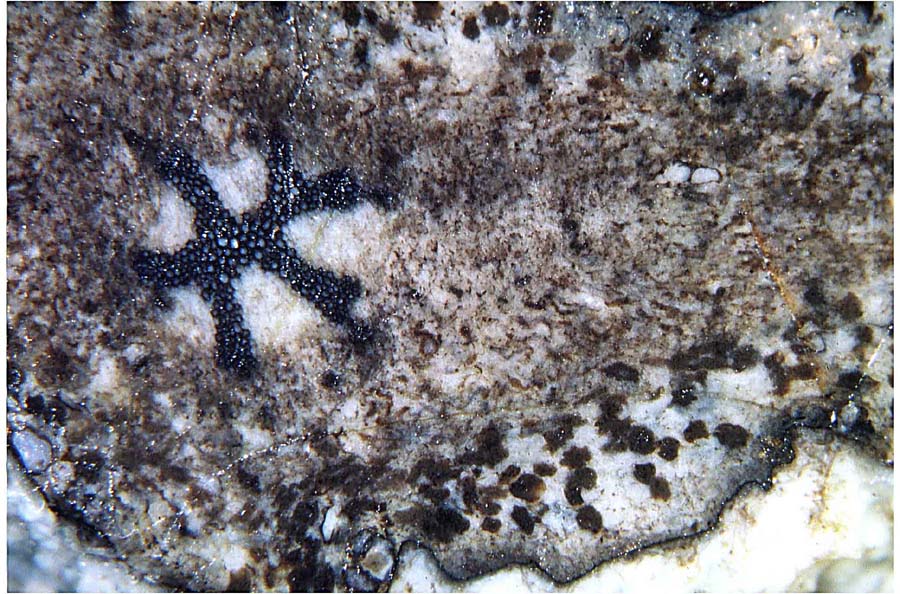
Fig.1: Detail of partially decayed Asteroxylon
cross section with ample evidence of fungus activity.
Shape and size of the angular
clots below right suggest their origin from individual cells, or
clusters of a few cells, filled with fungus matter. Image width 5mm.
Sample: Rh12/180, 0.12kg, 2007, Part 1.
The soft tissue of Asteroxylon
is nearly always severely damaged and squeezed. Patches of
tissue with preserved cell structure are
rare
exceptions, and so are the distinctly seen cells with dark content.
Most
probably the dark matter consists of a dense clot of fungus hyphae like
that of Glomites
in
Aglaophyton
cells, known as arbuscular mycorrhiza [1]. (A
remarkable image of clot formation by arbuscular mycorrhiza in Aglaophyton, with a
hypha seen penetrating the cell wall, is shown in [9], Fig. 19, and
[10], Fig. 3.96.)
Similar clots
in decaying tissue of Permian and Triassic plants have repeatedly been
interpreted as feacal pellets or coprolites of oribatid mites in
[4,5,6,7,8] and numerous other publications. (See Misconceptions.)
A few related images have been reproduced here: Figs.2-5.
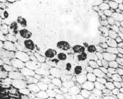
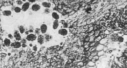
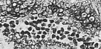
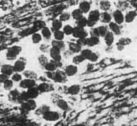
Fig.2,3,4 (right): Angular
clots in the tissue of the Permian
climbing fern Ankyropteris
brongniartii,
interpreted as mite coprolites in [4,5,6].
Fig.2: Sample Nr. K4568, Naturkunde-Museum Chemnitz. Width
0.63mm.
Figs.3,4: Sample Nr. K 4569, small
clots near tissue with small cells, larger clots near tissue with
larger cells. Widths 0.77mm, 1mm.
Fig.5 (far right):
Angular clots of various sizes and shapes in tissue with
cells of various sizes and shapes, alleged Triassic mite
coprolites [8], commented on here.
For a more detailed rejection of the elusive mites and their abundant
"coprolites" see Fossil
Wood News 23, 24 .
H.-J.
Weiss
2009, 2018.
[1] T.N. Taylor
et al.: Fossil arbuscular mycorrhizae from the Early
Devonian,
Mycologia
87(1995), 560-73.
[2] T.N. Taylor
et al.: Fungi from the Rhynie chert,
Trans. Roy. Soc. Edinburgh, Earth
Sciences 94(2004 for 2003), 457-73.
[3]
R.
Kidston, W.H. Lang: On Old Red Sandstone plants showing
structure, Part III,
Trans. Roy. Soc.
Edinburgh
52(1920), 643-680.
[4]
R. Rössler:
The late palaeozoic tree fern Psaronius - an
ecosystem unto itself,
Rev. Palaeobot. Palyn.
108(2000),
55-74.
[5] R. Rössler:
Der versteinerte Wald von
Chemnitz, 2001, p 141,155,169.
[6] R. Rössler:
Between Precious Inheritance and Immediate Experience,
in:
U. Dernbach,
W.D. Tidwell: Secrets of Petrified Plants, D’ORO,
2002, p 105.
[7] R. Rössler:
Two remarkable Permian petrified forests,
Geol.
Soc. London Special Publ. 265(2006), 39-63.
[8] D.W.
Kellog, E.L. Taylor: Evidence of oribatid mite detrivory
in Antarctica
during the Late
Paleozoic and Mesozoic,
J. of Paleontology
78(2004), 1146-53.
[9] H.
Kerp:
De Onder-Devonische Rhynie Chert ... , Grondboor
& Hamer 58(2004), 33-50.
[10] T.N. Taylor,
E.L. Taylor, M. Krings : Paleobotany,
Elsevier 2009.
 |
 |
28 |