Observations on fungus resting spores
 Resting
spores, often called by the not precisely defined term
"chlamydospores",
are formed separately on the fungus hyphae, probably with the aim to
persist when the hyphae decay. Since they are not comparable with the
plant spores formed closely together in capsules, the question arises
why they can be found in some places as densely spaced as in this
picture, where their volume fraction exceeds 1/2. From the aspect
of similar assemblages of silicified resting spores in the Rhynie chert
(Lower Devonian) it can be concluded that they had not been washed onto
the spot where they are seen now. They must have grown on the very
spot, and the hyphae as well as the decaying plant tissue which the
fungus had lived on must have vanished before silicification.
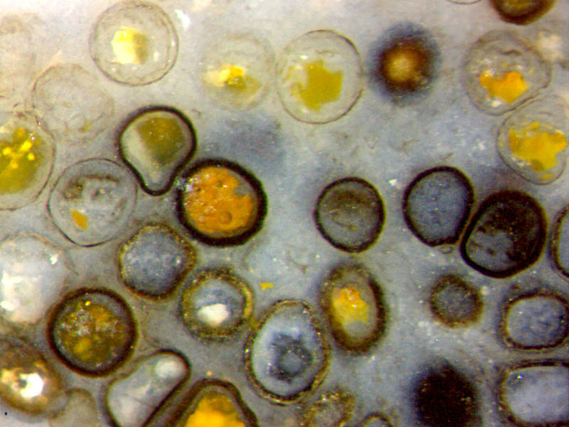
Resting
spores, often called by the not precisely defined term
"chlamydospores",
are formed separately on the fungus hyphae, probably with the aim to
persist when the hyphae decay. Since they are not comparable with the
plant spores formed closely together in capsules, the question arises
why they can be found in some places as densely spaced as in this
picture, where their volume fraction exceeds 1/2. From the aspect
of similar assemblages of silicified resting spores in the Rhynie chert
(Lower Devonian) it can be concluded that they had not been washed onto
the spot where they are seen now. They must have grown on the very
spot, and the hyphae as well as the decaying plant tissue which the
fungus had lived on must have vanished before silicification.
Fig.1: Assemblage of resting spores
(chlamydospores) of some fungus feeding on plant matter in the Devonian
habitat preserved as Rhynie chert. Width of the image 1.4mm.
How
the fungus did manage to produce this amount of organic substance would
be less problematic if the spheres consisted mainly of water, which
would make sense as a precaution against possible draught.
Most
peculiar is the highly different aspect of the spheres, considering
that probably they all belong to the same fungus species. Such
phenomena are known from neighbouring small cavities in volcanic rock
or in chert, also in the hollow aerial roots of Psaronius,
where tiny incidental differences in the chemical composition of the
enclosed or entering substances may cause widely differing developments
of the agate-like fills.
The separate ways of mineralisation
inside the spheres, resulting in different colours, are favoured by the
possible presence of a diffusion barrier around the sphere, which can
be deduced from Fig.2. The half sphere protruding from the raw surface
of the sample indicates that a crack running right against the sphere
had been deflected along its surface. Apparently the surface of the
fungus spheres is less well silicified so that the crack may use the
easy path around instead of right through. (The same is known from
terrestrial plants with their cuticle on the surface.)
Apparently
the crack deflection effect is a subtle one: More often the crack keeps
its plane path cutting through the sphere, as seen in Fig.2 on the
right.
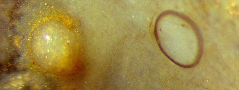
Fig.2: Rhynie chert, raw surface
with two resting spores bigger than those in Fig.1, the right one cut
through and the left one carved out by the running crack which had
separated the sample from the chert layer.
It
is not known why the spheres in one half of the assemblage in
Fig.1
have
got an apparently thick dark wall in the silicified state but are pale
and translucent in the other half, above left and beyond.
The thick dark walls suggest strength but three spheres left of the
middle indicate that things are more involved. One
sphere with thick
dark wall has become pear-shaped by contact while the two contacting
spheres have not become deformed at all. So it can be concluded that,
before silicification,
(1) the pale spheres can be as stiff as the thick-walled ones, and (2)
the latter can be less stiff than the pale ones.
Possibly the thick-looking wall
is not really thick but clad with a microbial layer which does not
contribute to stiffness.
This essay covers only a
small part of the questions related to the
several different resting spores corresponding to the several fungus
species found in the Rhynie chert, which "cannot yet be assigned to a
particular clade with certainty, as important parts of their life
cycles have not yet been discovered [1]." See also "Funny fossil
fungus formations".
H.-J.
Weiss
2016, 2017
[1]
T.N. Taylor,
E.L. Taylor, M. Krings: Paleobotany,
Elsevier 2009, 76.
 |
 |
104 |