Fossil fungus-related
spheres with alga connection
 Two preceding contributions
on fungus-related
spheres in one sample of Rhynie chert have provided surprising
observations. (See Rhynie
Chert News 116,
119.)
In the Rhynie chert in general, spheres and globules of any size
between about 10µm and 1mm are usually related
to fungi. They may be spores or fungus organs,
called vesicles or chlamydospores in case
of uncertain purpose, or sporangia,
zoosporangia, oogonia, and the bigger ones may be alga cells,
enormously bloated
under the influence of some chytrid fungus [1]. In view of
the different potential explanations, the interpretation of images like
these ones becomes quite interesting.
Two preceding contributions
on fungus-related
spheres in one sample of Rhynie chert have provided surprising
observations. (See Rhynie
Chert News 116,
119.)
In the Rhynie chert in general, spheres and globules of any size
between about 10µm and 1mm are usually related
to fungi. They may be spores or fungus organs,
called vesicles or chlamydospores in case
of uncertain purpose, or sporangia,
zoosporangia, oogonia, and the bigger ones may be alga cells,
enormously bloated
under the influence of some chytrid fungus [1]. In view of
the different potential explanations, the interpretation of images like
these ones becomes quite interesting.
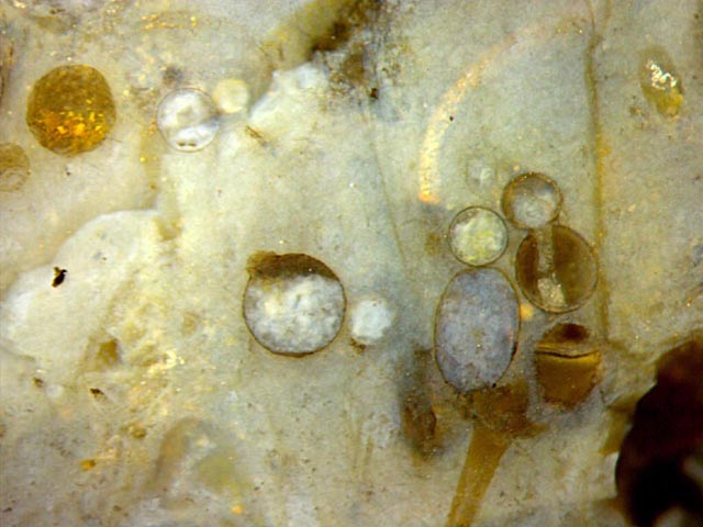
Fig.1: Cut plane of a small sample of
Rhynie chert with spheres up to 0.62mm, some of them arranged in a
peculiar way apparently related to a charophyte alga.
Picture width 4.3mm. All pictures: same scale, same sample as in Rhynie
Chert News 116.
Let us, for short, refer to the arrangement on the right of Fig.1 as
"aliens", one of them wielding a "sabre". The yellow sabre seems to be a
hollow
charophyte branch filled with reflecting quartz crystals. The lower
part of the left "alien" resembles an old charophyte whorl,
branches missing. For lack of another explanation, one may guess
that head and belly of the alien are bloated branch parts. This might
apply to the other alien, too: another charophyte branch transformed
into a still more strange chain of globules.
This guess is doubtful in view of the fact that there are no upper
branch parts emerging from the spheres, and that none of the other
spheres in this picture show any connection to an alga. This applies
also to the spheres, single or in pairs,
in the preceding contributions.
The assumption of a causal connection between
these spheres and a charophyte alga is supported
by early work on the Rhynie Chert [2]: Spheres
with sizes as seen in the present sample had been found
among branches of an alga named there Palaeonitella.
Moreover, some of the spheres were even found attached to the alga or
to another sphere, thus forming a pair. However, the authors [2] were
not right with their tentative interpretation of the spheres as "bulbils serving for vegetative reproduction".
 The origin of spheres and
globules of this type has convincingly been
explained by Taylor et al. [3] as alga cells bloated
under the influence of either of two fungi, Milleromyces and
Krispiromyces,
with characteristic features shown in great detail
[1,3,4,5]. A beautiful and instructive specimen of Palaeonitella with
two bloated internodal cells is shown in every one of these
publications, which might mislead to the assumption that this were
the usual arrangement of the globules. Hence the
numerous spheres shown in Rhynie
Chert News 116,
119
may serve as a useful reminder of the fact that spheres
or globules with sizes of about 0.3mm to 0.6mm are nearly always seen
not connected to an alga or its parts. Nevertheless
it is
uncertain whether or not most spheres within this size range had
originally been alga cells. An interpretation as fungus-affected alga
cells raises the problem of how they got the decay-resistant wall
distinctly seen here while the non-affected alga parts had mostly
vanished before silicification.
The origin of spheres and
globules of this type has convincingly been
explained by Taylor et al. [3] as alga cells bloated
under the influence of either of two fungi, Milleromyces and
Krispiromyces,
with characteristic features shown in great detail
[1,3,4,5]. A beautiful and instructive specimen of Palaeonitella with
two bloated internodal cells is shown in every one of these
publications, which might mislead to the assumption that this were
the usual arrangement of the globules. Hence the
numerous spheres shown in Rhynie
Chert News 116,
119
may serve as a useful reminder of the fact that spheres
or globules with sizes of about 0.3mm to 0.6mm are nearly always seen
not connected to an alga or its parts. Nevertheless
it is
uncertain whether or not most spheres within this size range had
originally been alga cells. An interpretation as fungus-affected alga
cells raises the problem of how they got the decay-resistant wall
distinctly seen here while the non-affected alga parts had mostly
vanished before silicification.
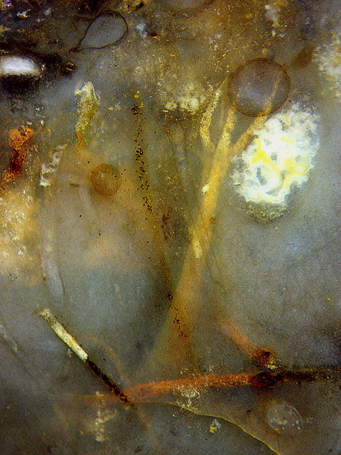
Fig.2: Rare sight of
several generations of Palaeonitella,
silicified in different ways: clear or filled with quartz grains, white
or stained with minerals; fungus-related spheres, 0.3mm and 0.6mm,
apparently not attached to the alga; swamp gas bubble
trapped among filamentous cyanobacteria, later filled with water and
silica by diffusion, then turned into white chalcedony. Image width
3.2mm.
Plain evidence for the presence of Palaeonitella
in this chert sample with abundant spheres is provided by Fig.2. It
shows an uncommon combination of several
successive generations of
this alga and two spheres preserved by silicification. Perhaps the
brown
specimen at the bottom and the big
three-pronged hollow fork with yellow fill in the background came next.
Two narrow tubes in front of the fork are incidentally aligned along
the cut
plane and thus seen despite of poor contrast. The three-pronged
fork in the background is probably hollow, with glittering quartz
crystals at the inner wall.
Doubts
concerning a close causal relation between spheres and algae are raised by Fig.3, with a sphere inside Rhynia. It is hard
to imagine
Palaeonitella penetrating into a dead plant and growing a
big sphere there.
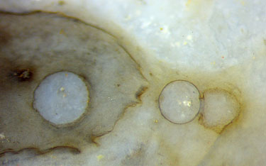
Fig.3 (right): Fungus-related
spheres, 0.5mm and 0.4mm, inside and outside Rhynia, apparently
not attached to alga parts. Image width 2.5mm.
Despite
of apparently contradictory evidence it may be concluded that probably
most of the big spheres or globules in the Rhynie chert are charophyte
alga cells hypertrophied and made persistent under the influence of
chytrid fungi in obscure ways.
H.-J. Weiss
2018
[1]
T.N.Taylor,
M. Krings, E.L. Taylor: Fossil
Fungi. Elsevier 2015, p.64.
[2] R.
Kidston, W.H. Lang:
On Old Red Sandstone plants … Part V,
Trans. Roy. Soc. Edinburgh 52 (1921), 855-902.
[3] T.N.
Taylor, H. Hass, W. Remy: Devonian
Fungi: Interactions with ... Palaeonitella.
Mycologia 84 (1992), 901-910.
[4] T.N.
Taylor, J.M. Osborn:
The importance of Fungi in shaping the paleoecosystem. Rev. Pal. Pal.
90 (1996), 249-262.
[5]
T.N.
Taylor, E.L. Taylor: The Rhynie
chert ecosystem: a model
for understanding fungal interactions,
in: Microbial Endophytes,
eds.:
Ch.W.
Bacon, J.F. White Jr., Marcel Dekker Inc., New York 2000.
For correction of contradictory data in [1,3,5] see here or
Google: errors palaeobotany.
 |
 |
120 |