Fungus-related fossil spheres in
Rhynie chert
The Rhynie chert, famous for its wealth of Lower
Devonian fossils, offers also spheres of various size, apparently most
of them at the end of some fungus hypha and known as chlamydospores
or resting spores,
possibly meant to survive unfavourable seasons.
They are easily recognized as such if the attached hypha is visible
(Fig.1) but most often the site of attachment is not in the picture
plane or the hypha had decayed before silicification (Fig.2). The
several different resting spores corresponding to the several fungus
species "cannot yet be assigned to a particular clade with
certainty, as important parts of their life cycles have not yet been
discovered"[1].
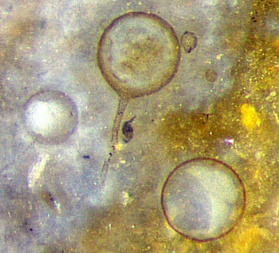
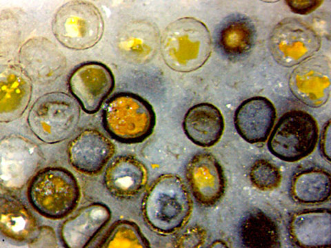
Fig.1 (far left): Chlamydospores of
some saprophytic fungus
among remains of early land plants.
Image width 0.6mm.
Fig.2: Chlamydospores of
some saprophytic fungus,
with stains obtained in silicification.
Image width 1.4mm.
Usually
the content of the chlamydospores had decayed
before silicification so that they appear empty or are filled with
deposits of different aspect unrelated to the fungus
species. From
mutual contact between the densely spaced fungus spheres in Fig.2 and
their
deformation it can be concluded that the spheres in Fig.2 had got
unequal
stiffness before silicification. The peculiar fact that the stiffness
of the sphere is not related to the
apparent thickness
of the wall is seen on the squeezed sphere in Fig.2
above left, which is deformed likewise by contact with the thin-walled
sphere on the left and the thick-walled sphere with red fill on the
right.
Much
less known than the several chlamydospores
are fungus-induced spheres whose generation
seems quite absurd: Under
the influence of a parasitic fungus a
charophyte alga miraculously grew big
spheres on thin
branches. For reasons unknown, the spheres persisted while the branches
decayed, which may mislead to the interpretation as big
chlamydospores.
Such danger of
misinterpretation reduces with the assumption based on observation that
interconnected
spheres
like those
in Fig.3 are always modified charophyte parts. The yellow spot with
three big spheres attached seems
to be the remains of a charophyte whorl of branches.
The spheres emerging from a charophyte
alga had probably been water-filled before silicification like the
chlamydospores mentioned above, and had become filled with mineral
deposits as known from small agates. There must
have been two fluid suspensions of dark particles, heavier
and lighter than the water inside the spheres. Either suspension
had separated itself
from the plain water by forming a plane horizontal boundary indicating
the orientation during silicification.
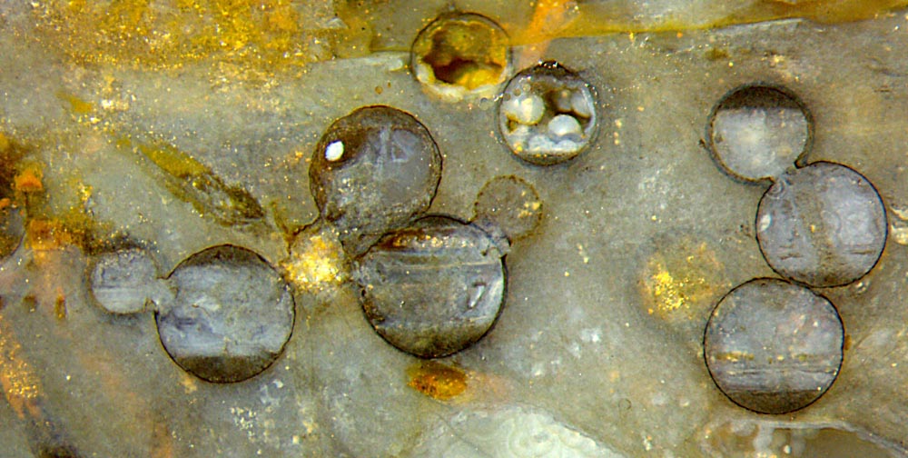
Apparently
the two smaller spheres above underwent another silicification regime
with silica concentration so low that the content did not silicify as a whole but silica became deposited on the walls, thereby forming
spherolites resembling glossy pearls.
Fig.3:
Well-formed spheres resulting from malformed growth of
the charophyte
alga Palaeonitella
under the influence of the parasitic fungus Milleromyces [2].
Image width 5mm.
One may wonder why fungi produced nearly perfect spheres in
quite different ways:
(1) chlamydospores as persistent organs at the ends
of hyphae, (2) hypertrophied
alga tubes due to the
parasitic fungus Milleromyces
interfering with the normal growth
of a charophyte [2].
Fig.1: Rh2/29.4, obtained
from Shanks in 2001; Fig.2: Rh12/160.9, found
in 2007; Fig.3: Rh14_17.5, obtained from
Barron in 2007.
H.-J. Weiss
2022
[1] T.N. Taylor,
E.L. Taylor, M. Krings: Paleobotany, Elsevier
2009, p76.
[2] T.N.
Taylor, M. Krings, E.L. Taylor: Fossil
Fungi. Elsevier 2015, p64.
 |
 |
190 |