Devonian fungus globules densely
clustered
Globular organs of several fungi
are often seen in the Lower Devonian Rhynie chert. They may be conspicuous,
scarce or abundant,
and randomly distributed in
some places inside or outside plant tissue. 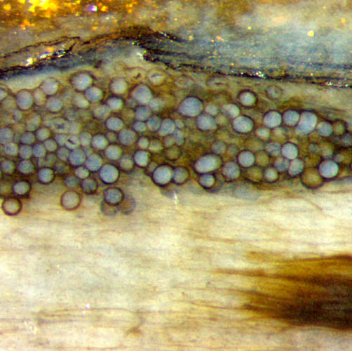
The pictures, taken from three samples, are of equal scale
and width of
1mm. They show fungus globules that are densely packed for
unknown reasons.
Fig.1: Coherent cluster of fungus globules, 35-40µm, adhering to the
surface of Rhynia.
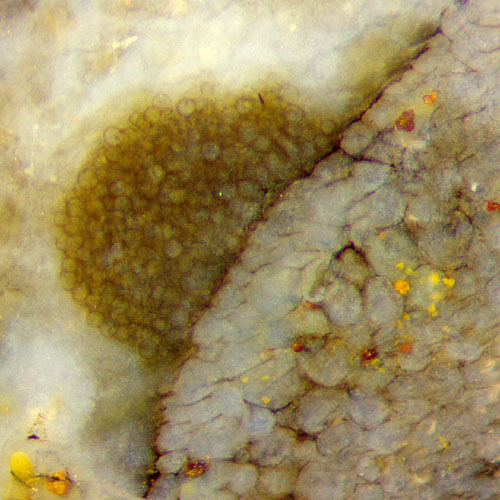
Fig.2 (far right): Cluster of fungus
globules, 40-60µm, inside degraded Rhynia.
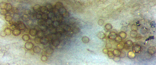
Fig.3 (below): Two clusters
of fungus globules, 35-50µm, in an area of microbial filaments and networks.
Fungus
hyphae are known to thrive inside or near decaying plant matter or even
within live plants. They may form vesicles, mostly more or less
spherical, often called globules or
chlamydospores, a not well defined term. The clusters of fungus globules
in these pictures give rise to wonder.
For
no obvious reason, they may stick together closely, as in Fig.1, where
they form a lump adhering to the surface of Rhynia which does not show
signs of decay. Possibly they are kept in place by hyphae not
seen here but what is the advantage of clinging to each other and to a
plant that is not decaying ?
Even inside the decaying plant in Fig.2 it remains enigmatic why the
hyphae have grown their globules huddled together.
 The
globules in Fig.3 have assembled in an area where there is no plant
part in the vicinity, only large swaths of filiform and net-like
microbes. Again the question arises what the fungus had been feeding on
before it became able to grow clusters of spheres.
The
globules in Fig.3 have assembled in an area where there is no plant
part in the vicinity, only large swaths of filiform and net-like
microbes. Again the question arises what the fungus had been feeding on
before it became able to grow clusters of spheres.
As
a confusing peculiarity, the clusters of this fungus, looked at with
moderate magnification as in these cases, may resemble heaps of spores.
This visual impression is brought about by apparent markings faintly
seen on some of the globules similar to the trilete marks on spores or
to the aspect of spore tetrads. These are illusions due to the mostly
high transparency of the spheres, as seen, for example, on the sphere
at the right edge of Fig.3, where the outlines of two or three spheres
in the depth are faintly seen through.
The same effect is also apparent with a picture of Zwergimyces
in the latest monograph on fossil fungi [1]. This effect disappears
with higher magnification, where the spheres show lots of
detail,
including hyphae and a mantle of complex structure [2], but not the
illusory markings.
The
similarity of this Fig.1 and Fig.6.9 in [1] seems to justify the
assumption that the present pictures taken from three samples of Rhynie
chert show Zwergimyces,
even though Fig.1 and Fig.3 do not support the claim in [1] that
"Zwergimyces ...
occurs in degraded plant axes ...". Another
example of clustering transparent spheres like the present ones has
been seen in a microbial
lump which apparently had solidified into a miniature reef in
the Devonian swamp at Rhynie.
The questions raised above, concerning the reason for the close
clustering of globules, also in areas without decaying plants, have not
been considered in [1,2]. The possible affiliation of Zwergimyces has
remained an unresolved problem, which is discussed in detail in
[2].
H.-J. Weiss 2017
[1] T.N.Taylor, M. Krings, E.L. Taylor:
Fossil Fungi, Elsevier 2015, p.80.
[2] M. Krings, T.N.Taylor, N. Dotzler, C.J. Harper: Morphology
and ontogenetic development of Zwergimyces
vestitus ...
from the Lower Devonian Rhynie chert.
Rev. Palaeobot. Palyn. 228(2016), 47-56.
 |
 |
115 |