Fossil fungus-related
spheres that come in pairs
Spheres
related to fungi
are a common sight in the
Rhynie chert. Usually they are separate and randomly distributed but
the small spheres of one species come in dense
clusters. Quite uncommon
is the phenomenon of big spheres in pairs. Considering that the cut
would not show connected spheres unless their position and orientation
with respect to the cut plane is within narrow limits, it may be
concluded from
Fig.1 that most of the spheres are arranged in pairs here but not all
are seen so.
There
is no obvious reason for this pair formation. The problem will not be
resolved here but by looking at the details one may be guided along the
way towards an explanation.
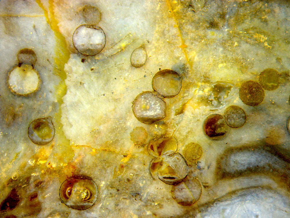
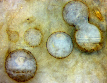
Fig.1: Cut plane of a small sample of
Rhynie chert with fungus- related spheres up to 0.7mm, mostly
arranged in pairs, connection not always in the cut plane. Image width
5.54mm.
A phenomenon not related to the fungus connection but nevertheless
interesting is the formation of dark deposits at the bottom and /or at
the top of the spherical cavities. Apparently there had been different
suspensions (of mineral precipitates, dead microbes etc.) in water that
were either heavier or lighter than water and settled accordingly.
Additional evidence for the conspicuous pair
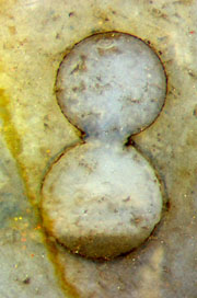
formation is provided by Figs.2-6. Level deposits of suspensions are
seen in Figs.2-5.
The
deposit in Fig.5 consists of a stack of layers, which indicates that
the deposition took some time with periodically changing conditions.
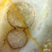
The highly symmetrical aspect of the fused spheres in Fig.2
indicates that (1) the spheres are of nearly equal size, (2)
incidentally the axis of rotational symmetry is well within the cut
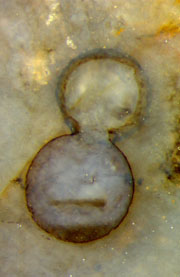
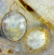
plane. For comparison, the axis is slightly inclined to the cut plane
in Fig.3, where the upper sphere is slightly larger than its circular
section on the cut face.
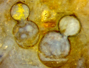
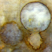
Rather well symmetrical cuts, except for the sphere
size, are seen in Figs.4,6, each one with a remarkable detail: There is
a small stub, possibly a broken-off or budding hypha, on the smaller
sphere close to the symmetry
axis.
Figs.2-6: Fused spheres of equal or moderately differing sizes.
Same scale as above, width of the
pictures 1 or 2 mm.
Figs.7,8
(below): Spheres with outgrowths. Picture width 1mm. All pictures have
been taken from the same chert sample, same scale.
The seemingly clear principle prevailing in this peculiar sample of
Rhynie chert, which is fungus-related spheres fused into dumbbells,
becomes less clear in several ways.
It
is not clear whether the outgrowths in
Figs.7,8 become as large and round like another sphere, as possibly
indicated by two "would-be dumbbells" in Fig.1 (above left). Dark dots
are distinctly seen on the surface of spheres excentrically cut, as in
Figs. 5,7.
A small globule in Fig.5 (above left)
seems to be germinating, thereby producing a 50µm-tube
with a bulging end of 85µm.
As a disturbing fact, charophyte tubes and whorls, probably Palaeonitella,
are present in this sample, often quite near to these spheres, as in
Fig.7, where a tube is partially filled with a dark former suspension.
Since it is known that charophyte tubes can be induced to grow big
globules under the influence of fungi [1], the suspicion arises that
the spheres seen here might be bloated charophyte cells like
those in [1] but about twice as big. (See Rhynie
Chert News
119,
120.
)
However, the 40µm-tubes seen as stubs in Figs.4,6
and elsewhere are much smaller than the charophyte tubes of
90µm - 300µm in
this sample but compatible with big fungus tubes of 35µm in this sample
so that an interpretation as expanded fungus hyphae or
chlamydospores rather than charophyte cells seems appropriate.
Large
spheres with diameters up to 0.75mm and more, some
with adhering hypha, have been found in
several samples but this sample has been the only one with
dumbbell-like formations.
Further details are expected from other cut planes of this sample.
Sample: Rh14/17 (0.35kg), obtained from Barron in
2007.
H.-J.
Weiss 2017, 2018
[1]
T.N.Taylor,
M. Krings, E.L. Taylor: Fossil
Fungi. Elsevier 2015, p.64.
 |
 |
116 |