Fossil fungus-related spheres with fancy fills
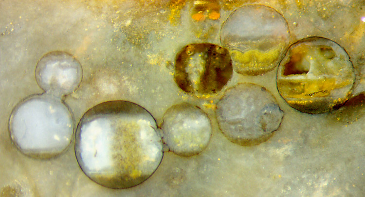
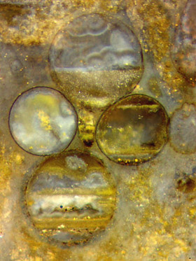
Fig.1
(far left): Spherical former water-filled cavities of dubious origin,
some pairwise fused, partially still hollow but mostly filled
with chalzedony from siliceous
precipitates forming clouds or deposits with level boundaries.
Picture width
3.5mm.
Fig.2: Similar as Fig.1, chalzedony grown as level layers or as clouds,
governed by changing silicification regimes.
Picture width 1.3mm.
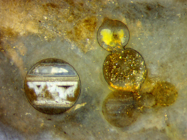
Fig.3
(below): Similar as Figs.1,2, complex and
distinctly different silicification products
inside spheres, with silica grain sizes covering the range
of light wavelength (bluish), reflecting grains (white), and coarse
quartz crystals (glittering).
Picture width 2.8mm.
 Fossil
spheres in chert, incidentally cut right through the middle, are often
the most conspicuous objects on the cut face
owing to a thin black circular line. Hence, a thin surface layer of the
sphere must have persisted while the innards,
like the tissue
of nearby plants, had largely decayed before the sphere became filled
with
various deposits.
Fossil
spheres in chert, incidentally cut right through the middle, are often
the most conspicuous objects on the cut face
owing to a thin black circular line. Hence, a thin surface layer of the
sphere must have persisted while the innards,
like the tissue
of nearby plants, had largely decayed before the sphere became filled
with
various deposits.
So
one
is tempted to assume that the spheres are fungus organs known as
chlamydospores or resting spores from numerous fungus
species, and
that after the decay of the fungus substance inside, silicification
processes driven under varying conditions by silica gradually enterig
by diffusion produced the fills.
However, this seemingly straightforward logic is subjected to doubt by
the rather large size of the spheres and by observations
hinting
at a quite different possible explanation as hypertrophied alga cells,
as considered
before.
This makes the fascination of dealing with the phenomenon. Regardless
of what might be revealed by further investigations, let us
consider the fills here.
Most former cavities
in cherts formed by
silicification of swamp matter or water had been swamp gas bubbles
trapped among plant debris or microbial slime. They differ from the
spheres in these figures by their irregular shape. After
the gas had escaped and water had come in by diffusion through the
silica gel, they were just water-filled cavities ready for subsequent
processes of mineralisation and deposition, not much different from the
spheres considered here after the decay of their organic content.
In the water-filled cavities of either type, precipitates
or suspensions may form, which most often settle at the bottom. Less
often they settle at the top of the cavity, which means they are
lighter than water, which can be expected from dead microbes or alga
unicells, for
example. Some of the spheres in these
images have got dark deposits at
both top and bottom. The boundaries of the deposits formed from
suspensions, of course, indicate the horizontal direction during
silicification.
It
can be deduced from the extremely different aspect of fills in
any
of these figures that even minor differences in the chemical
composition of neighbouring cavities or even
of compartments inside the same cavity can strongly influence the
result of
silicification.
In Fig.2, upper sphere, a fine-grained suspension had settled at the
bottom, then silica gel of cloud-like aspect
had formed in the water above.
In Fig.3, big sphere, a highly complex silicification regime must have
been at work.
Certainly the dark deposit of light suspension at the top and the gray
horizontal plate must have been there when the bluish fill
formed
in between. The structure below the gray plate is rather confusing,
apparently a result of dissolution of gel after the plate had formed,
then governed by gel formation again. (Alternating formation and
partial dissolution of
silica gel is not uncommon during chert formation.)
On the right in Fig.3, the result of silicification differs much
between
the two spheres even though they are connected
by a wide neck. Apparently, the formation of gel and chalzedony in the
upper compartment
had used up nearly all silica so that in the water below, quartz
crystals grew slowly at a pace restricted by the influx of silica
from outside via slow diffusion.
Same sample as in Rhynie Chert News 116.
H.-J.
Weiss 2017
 |
 |
119 |