Silicification steps in chert deduced
from fossil fungus hyphae
Chalzedony differing locally in hue and brightness
allows a complex sequence of silicification stages to be reconstructed
from Fig.1. This is made easier and more convincing by the presence of
fungus hyphae which obviously became covered with a whitish coating
which
contrasts against a dark brown matrix. Hyphae
found outside plant
matter must have grown and become silicified
in water. As another conspicuous feature, the
stack of levels resembling agates with horizontal banding indicates
discontinuous silicification, which will be explained below. The
sequence of events which materialized on the small area on
the cut face
of the Rhynie chert sample shown here begins with
a former swamp gas bubble trapped among plant parts.
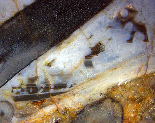
Fig.1:
Complex-structured fill within a former
swamp gas bubble in Rhynie chert.
(Lengthwise section of
the early land plant Aglaophyton
above left.) Note the level deposits,
slightly tilted towards each other.
Width of the image 10mm.
Fig.2: Detail of Fig.1. Note the hyphae of some aquatic
fungus coated with
whitish chalzedony whose thickness varies with height above bottom.
Width of the image 4mm.
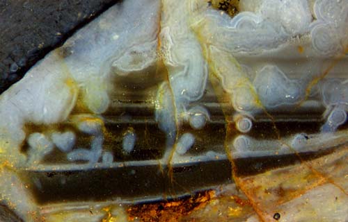
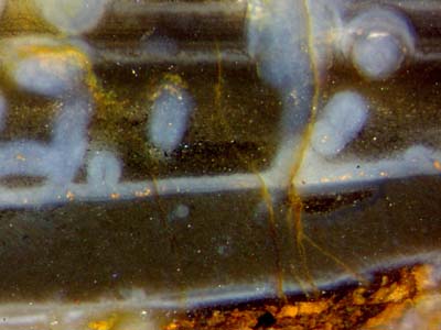
Fig.3 (on the right): Detail of
Figs.1,2. Note the two small cross-sections
of pale coated fungus
hyphae, diameter about 40µm, in the lower dark stratum. Width of the
image 1.6mm.
The apparent succession of events which brought about what
is seen in Fig.1 and details thereof is listed here:
(1) A patch of Lower Devonian vegetation became
upset and inundated in silica-rich water.
(2) A swamp gas bubble got trapped among plant parts.
(3) The silica solution became supersaturated and
turned into silica
gel rapidly ere the plants could
rot.
(4) With silica gel formation, the gas bubble became stabilized.
(5) After this early silica deposition had essentially ended, the gas
slowly escaped by diffusion
through the surrounding gel while water
entered in the same way.
(6) Other than the surrounding gel, the now
water-filled cavity provided a habitat for some aquatic
fungus to thrive. A
loose tangle of fungus hyphae spread throughout the water.
(7) Much later than the light-weight and hence quickly moving water
molecules, the heavier silica molecules or
complexes entered by diffusion and slowly increased the
silica concentration there.
(8) Apparently the
solution became supersaturated again, only slightly since the
water did not turn into gel as it did before, see (3).
(9) The silica complexes or clusters were still
so small that they got pushed about by thermal motion such that their
concentration remained homogeneous throughout
the water. Silica deposition began with the formation of a thin white
coating on available substrates, that is the cavity walls and
the
fungus hyphae. The result of this first silicification step inside the
water-filled cavity is seen as a thin white layer and as coatings of
about 20µm in the lowermost part. (The hyphae are so thin that the
diameter
of the coated hypha equals twice the coating thickness.) Two
cross-sections of
coated hyphae are vaguely seen in Fig.3 as small pale spots.
(10)
Apparently, as the silica clusters became larger, they settled to form
a comparatively heavy suspension
with a well-defined boundary against the lighter solution above. This
must have been a fast process since the
diameter of the upper coated hypha is not
larger than that of the one at the bottom of the fill: If the
level had risen more slowly, the upper hypha would
have acquired a thicker coating while it was not yet immersed in the
settled suspension. The dark stain of the suspension could possibly be
due to the
decay products of microbes living in the water.
(11) The accumulation of
dark suspension stopped. It solidified and became the lower
stratum of dark silica gel, now chalcedony, in Fig.1.
(12) Again with slight supersaturation, the process described in (9)
repeated itself in the cavity of reduced size. The whitish lining and
coatings are obviously much thicker this time.
This means that their deposition went on for some time without being
interfered by new-formed dark suspension.
(13) Eventually, suspension
formation as described in (10) repeated itself at a slower pace. The
rising level stopped the growth of the whitish lining and
hypha coatings as soon as it reached them.
(14) The level of settled suspension rose so slowly this time that
there was
more time available for the growth of coatings
on the hyphae higher up. This is evident in Fig.2 where the diameter of
coated hyphae is 130µm below and 200µm above in
the second dark stratum, which is bounded by the next white line whose
origin is explained below.
(15) Meanwhile the solidifying swamp had got a slight tilt. The
new interface level has been choosen as the horizontal
direction in these pictures so that the old level is slightly tilted
against the new
one.
(16) Again, the accumulation of dark suspension stopped. Same as before with the lowermost stratum,
it solidified and became the second stratum of dark silica
gel.
(17) In the silica solution above, a third lining
of whitish silica gel was deposited around the new cavity walls, and
since three coated hyphae, seen in Fig.2 as cross-sections, partially
stuck out, they, too, became covered by the third white
lining. This is a peculiar type of coating: lower half
thin, upper half thick.
(18) A third cycle of accumulation of dark suspension ended the deposition of white
gel again.
(19) Same as described in (16), a third dark stratum was
formed.
(20)
All the time while the lower strata were formed and solidified, the
hyphae higher up in the water-filled cavity surrounded themselves with
ever thicker coatings which fused into a coherent mass of white gel
with a few pockets of water in between.
(21) Eventually, suspension
formation and solidification ended further deposition of white gel
there, too. By that time, the thickest coating had reached a diameter
of 270µm, hence a diameter of the coated hypha of 540µm, as seen in
Fig.1 on top.
The succession of processes which led to the agate-like fill and coated
fungus hyphae in this former
cavity may be confusing so that a summary is appropriate: Watery
suspensions of silica clusters segregate if the particles are so big
that they settle into a fluid precipitate with a horizontal
interface against the water above. Any horizontal
plane faces in cherts, often thought to be former
water levels, are former interfaces between fluids.
They could not have been former water levels since mm-size
water surfaces against air would be spherical bubbles.
Successively
formed suspensions may vary in aspect but in the present case it is
always the same dark brown, the cause of which can be guessed and may
be foumd out with higher magnification.
The white precipitate is not a settled suspension
since it appears as hypha coatings, too. Plane white
layers
could
have been deposited only with the brown suspension below being in a
solid state, and without new brown particles being formed. Hence, the
production of dark suspension
must repeatedly have stopped for a while, then started anew later.
Similar discontinuities in
silicification are well known from banded agates, where the
"bands" seem to have been deposited separately.
From the sorting of the coated hyphae according to diameter,
with thicker coatings positioned higher up than thinner ones, it can be
concluded that hypha growth was restricted to an early time span when
the silica concentration in the water-filled cavity was so low
that no coating was formed. Otherwise, if new hyphae had grown
in
the
later stages, too, no relation between coating
diameter and height above the bottom of the former cavity would be
observed.
Finally
it can be stated that the Rhynie chert sample shown here, with whitish
coatings and dark horizontal strata, offers evidence of discontinuous
silicification whose cause remains obscure.
Sample: Rh12/91, (0.31kg, Part5: slab 6-7mm),
found near
Smithston in 2006.
H.-J. Weiss
2015,
2016, 2018
 |
 |
77 |