Symbiotic fungus in Trichopherophyton ?
Cells with dark fill, seen on cross-sections
of Aglaophyton
(former
Rhynia major)
as a dark ring at some distance below the
epidermis, is a common sight in the Lower Devonian Rhynie chert.
Quite similar rings on cross-sections of the smaller Rhynia
are not rare. The phenomenon has been
described in detail and explained as being due to the fungus Glomites rhyniensis
engaged in a type of symbiosis known as
arbuscular mycorrhiza [1]. It would
be interesting to know, of course, whether or not such symbiosis had
been more widespread among the Lower Devonian plant species.
Evidence is provided here of the occasional
occurrence of quite
similar arrangements of cells with dark fill in the "hair-bearing
plant" Trichopherophyton,
which is not related to Aglaophyton.
It is
one of the less abundant plants in the chert, and it is easily
recognized only if the less often preserved upper parts with the
bristles are seen, as in these pictures. (The
big bristle below left in Fig.1, length 1.55mm, might be one of the
longest ever seen on this plant. It can easily
be mistaken for a crack.)
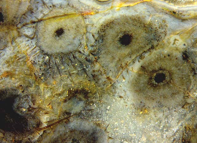
Fig.1 (left): Three bristly
cross-sections of Trichopherophyton
in
full view and about five more partially seen on this small
area of 7mm width. Note also the big bristle below left.
Figs.2,3: Cells with dark fill loosely arranged as a dark ring on Trichopherophyton
cross-sections.
Width of the pictures 4mm. Same scale for Figs.1-3.
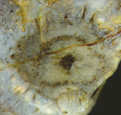
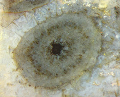
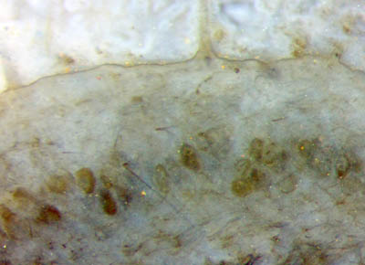
Fig.4 (below): Detail of Fig.3.
Cells with dark fill and
definite shape
contrasting to the degraded tissue around.
Width of the picture 1.3mm.
Apparently
the plant parts with dark cells as those in Figs.2,3 and others had not
been less vigorous than those without. Thus one may conclude that
the dark fill did not do any harm and perhaps had even been beneficial
as previously assumed in connection with Aglaophyton [1].
As seen in Fig.4, the dark fill is confined to individual cells which
are in a better state of preservation than the largely decayed tissue
harbouring them. The
hyphae vaguely seen in Fig.4 as thin dark lines could as well belong to
some rot fungus which entered into the dead plant.
All pictures taken from Sample Rh14/18, obtained from Barron in 2007, Part 4 (slab).
What has to be done with higher resolution is to show that
the dark fill is made up of fungus arbuscules. This would be another piece of evidence
of this type of symbiosis between fungi and early land plants.
H.-J. Weiss
2015,
2018
[1] T.N. Taylor
et al.: Fossil arbuscular mycorrhizae from the Early
Devonian,
Mycologia 87(1995), 560-73.
 |
 |
76 |