Devonian gloss
Vegetable cell surfaces keeping glossy for several hundred million years are
worth being noticed. In the following, two quite different occasions of
large Devonian cells having kept their original gloss are presented. They may have lain protected but unnoticed in chert until a
crack revealed their presence, as in the below examples of fungus
spheres, or the protecting chert is so transparent that the
glossy
face is seen without being exposed, as in the below example of a
charophyte alga.
Chlamydospores
or resting spores are produced by fungus hyphae locally expanding into persistent spheres. They are common fossils
in the Lower Devonian Rhynie
chert [1] but seldom seen as numerous and beautiful as on the
rather smooth 3mm-wide patch of crack
face in Fig.1 which is part of the sample surface now.
Apparently the thoroughly silicified
globules did not act as inhomogeneities so that the crack went right
through, randomly cutting them or passing them by so that some are
still seen as whole spheres in the depth below the crack face. The
conspicuously variable aspect
of the globules in this picture has been discussed before in Rhynie
Chert News 104,
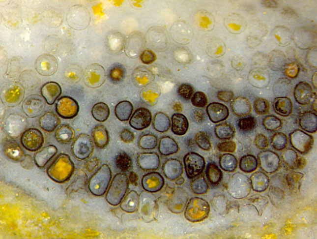
Fig.1:
Uncommon assemblage of persistent fungus resting spores in a decayed early
land plant in Rhynie chert, hyphae decayed and vanished. Picture taken on the natural
surface of the chert sample. Image width 3mm.
Accumulations of resting spores as in Fig.1 are exceptional. Usually
they are seen scattered on the surface or on cut
faces of the chert samples. A few of them make beautiful pictures:
Rhynie
Chert News 163
.
A rare sight are spheres protruding from a
fracture face, as seen in the pictures below. Apparently
the crack
heading for an inclusion may get deflected around it. As a
lucky coincidence, the
two options of crack propagation near a globular inclusion,
straight through or around,
are seen realized by the same crack in the same material:
Fig.2. As one may guess from Fig.1, the
dark or clear surface aspect in Fig.2 is due to a secondary phenomenon,
like the
presence or absence of microbial intruders, and therefore irrelevant
here. This may also apply to the different aspect of Figs.3,4.
The
comparison with Fig.1 may be justified although Figs.2-5 show other
samples with larger globules (except Fig.5). Note that the scale of
Figs.2-5 is twice that of Fig.1. All these globules partially laid bare
by propagating cracks in the disintegrating chert layer were found on
the surface of chert samples. No attempt is
made here to relate these resting spores
to certain fungus species in the Rhynie chert.
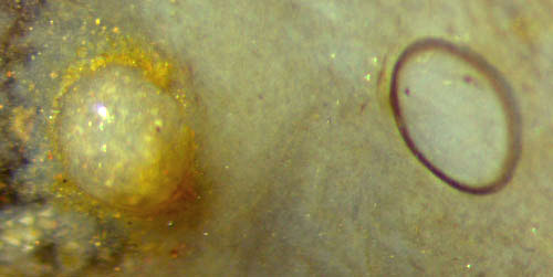
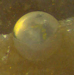
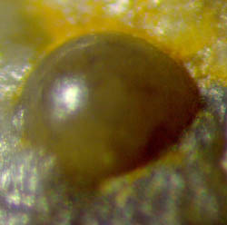
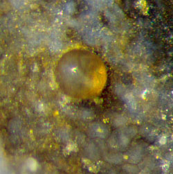
Fig.2: Two fungus globules in the Rhynie chert laid bare by a crack
propagating right through one of them and along the surface
of the other one. Image width 1.2mm.
Fig.3: Rather perfect sphere protruding from the sample
surface. Diameter 0.39mm.
Fig.4: Dark sphere with small deviations from perfect shape revealed by
slightly fuzzy reflection. Diameter 0.44mm.
Fig.5: Sphere comparable to the clear ones in Fig.1. Diameter 0.175mm.

The
glossy reflections in Figs.2-5 indicate that the globular faces,
after having been laid bare by propagating cracks, were not affected by
subsequent mineral or microbial deposition. Doubtless unaffected is the
alga enclosed in silica gel, now clear chalcedony, in Fig.6
since it had never
been laid open. The incident light entering into the clear
chalcedony gets partially reflected at the alga tube (which consists of
one cell). This reflected
part, after traversing the clear
chalcedony towards the observer, is seen as gloss. That part of the
incident light which enters into the alga tube traverses the tube
content and gets
partially reflected at the rear wall of the tube.
It reaches the observer after having twice traversed the outer
chalcedony, twice the tube wall
with interface, twice the tube content, and having been reflected at
the back
of the tube. The light remaining after these 7 successive losses of
intensity is still sufficient for a faint gloss at the inner tube
boundary seen
beside the main gloss in Fig.6. See also Rhynie
Chert News 74
.
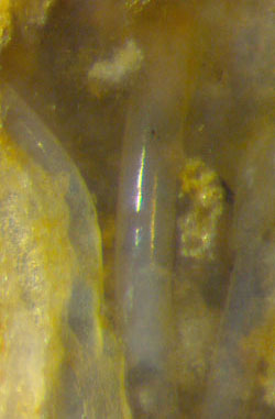
Fig.6: Devonian charophyte alga stem filled and embedded with clear
chalcedony. Image width 0.6mm.
Fig.7: Devonian charophyte alga stem in a cavity, coated with quartz.
Image width 0.3mm. Same scale for Figs.2-7.
Mirror-like
reflections indicate that the reflecting face is smooth on a sub-µm
scale. Watery solutions with high supersaturation of silica make silica
gel which protects
the smoothness: Fig.6. Low supersaturation gives rise
to slow formation of tiny quartz crystals and their deposition on the
once glossy
surface, as seen with another alga stem in a once water-filled but now
empty cavity in the same chert sample: Fig.7.
The
different aspects of the same alga species surrounded by
chalcedony (Fig.6) or by air (Fig.7) are mainly due to
light scattering. The chalcedony inside and outside the tube in Fig.6
is essentially transparent, hence it is inconspicuous except for the
gloss. The alga coated with tiny quartz crystals in Fig.7 scatters and
traps the incident light so that it appears brighter than the one with
perfectly preserved surface in Fig.6.
Samples (weights refer to the whole sample):
Fig.1: Rh12/160.9 (0.54kg) found in 2007; Fig.2:
Rh2/226.2 (32g) found by S.W. in 2014;
Fig.3: Rh2/166.1 (0.36kg) obtained from Shanks in 2011;
Fig.4: Rh12/166.1 (45g) found in 2006;
Fig.5: Rh2/354 (62g) found by S.W. in 2014;
Fig.6,7: Rh5/3.2B,3.2A (1.5kg) found by S.W. in 2001;
H.-J. Weiss 2021
[1] T.N.Taylor, M. Krings, E.L. Taylor: Fossil Fungi, Elsevier 2015.
 |
 |
177 |