Common fossils with uncommon aspect in
the Rhynie chert
The
famous Rhynie chert fossils representing a Lower Devonian ecosystem
have become widely known by publications in monographs and textbooks.
Less well known but all the more interesting are the occasionally
encountered deviations from the commonly published forms. As a
remarkable fact, the following examples of less common structures have
been found together on one Rhynie chert slab.
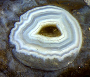
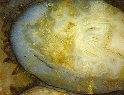
From a botanical point of view, the introductory picture
of Fig.1 is the most unproblematic one. It is hollow Aglaophyton with
everything decayed except for the cuticle on the former epidermis,
which had defined the cavity for the agate to be deposited in the same
way as agates are usually deposited in silica-rich water.
Fig.1: Agate formed in a cavity left after the decay of Aglaophyton
lying prostrate in the swamp water. Image width 3mm.
Fig.2 (right): Aglaophyton
with preserved tissue but central strand missing, with uncommon
radial fissures and shrivelled surface.
Image width 4mm. Same
scale for all figures.
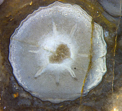
Radial fissures like these, emerging from a destroyed central strand,
have not been seen on thousands of Aglaophyton
cross-sections on own samples. They clearly differ from the conspicuous
fissures apparently
related to fungus infection [1].
Fig.3 (below left): Aglaophyton
cross-section with
rare type of damage above and a horizontal
layer below, resulting
from a dark precipitate deposited at an early stage of
silicification. Image width 4mm.
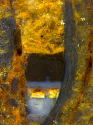
Fig.4 (below right): Stack of horizontal layers deposited
successively with different aspect for reasons unknown.
Image width 3mm.
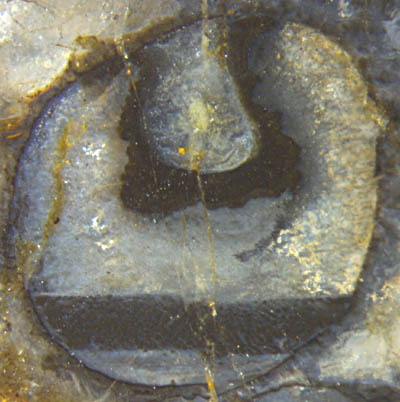
The horizontal deposits
seem to be formed by precipitation of tiny particles making a fluid
suspension before everything turns into chalcedony. The thick black
coating around the hole in Fig.3 remains unexplained here.
Irregularities in the
yellow layer in Fig.4 indicate that there had been hyphae coated with
bluish silica gel when the yellow suspension settled.
Fig.5 (below): Aglaophyton,
inclined cross-section with
remains of tissue (bluish), former inner cavity with fungus
hyphae, and microbial
formations outside.
Image width 4.2mm.
The
hyphae in Fig.5 had grown in the water-filled cavity and got surrounded
by a thick coating of pale silica gel. Remarkable are the wavy hyphae
among the perfectly straight ones. The
wavy hyphae might be the mycoparasite Trichoderma.
The irregular or pointed shapes grown on the surface resemble those
known from the blue-green alga Croftalania.
The combination of wavy hyphae and Croftalania
has been observed here for the first time.
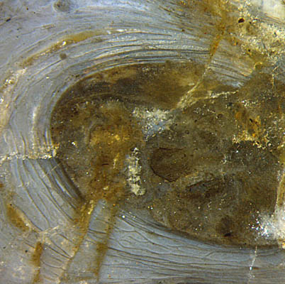 Fig.6 (left): Aglaophyton
with thin microbial sheets wrapped around,
inclined cross-section. Image width 4mm.
Fig.6 (left): Aglaophyton
with thin microbial sheets wrapped around,
inclined cross-section. Image width 4mm.
Stacks of thin microbial
sheets [2]
are not rare in the Permian cherts from the Döhlen basin
(Saxony) but seem to be rare in the Rhynie chert
[3].
Apparently, microbial sheets wrapped
around plants like those in Fig.6 have not been seen before. Their
branching and merging indicate
a way of formation different from that of the Permian sheets in [2].
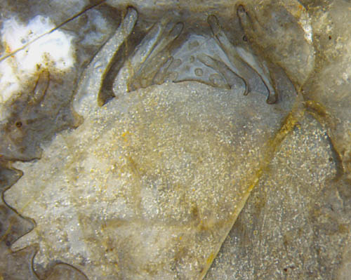 Fig.7 (right): Peculiar
structure suggesting an unfolding bud.
Fig.7 (right): Peculiar
structure suggesting an unfolding bud.
Image width 5mm.
The fossil in Fig.7 would readily be recognized as Asteroxylon if
there were not the intriguing fact that none of the several cut faces
of this big chert sample shows the characteristic features,
the complex-shaped
cross-sections of the central strands, which usually
reveal the presence of Asteroxylon
even if poorly preserved. The large number of Asteroxylon sections
shown in [4], none of them resembling this one, give rise to the
suspision that the object
in Fig.7 might be something special. It does not appear here as the top
of a larger
plant but seems to be a peculiar blob of tissue of its own, with a kind
of broad bud just
unfolding.
All of the grown-up plants in this sample, like the ones in the above
figures, are
lying prostrate along the chert layer, apparently upset by a sudden flow,
with only the
little lump oriented such that it suggests upward
growth. While the tiny leaf-like forms on top strongly resemble the big
enations of grown-up Asteroxylon,
the distinctly different lateral protrusions on the left are
problematic. In view of the fact that the gametophyte of
Asteroxylon
has not yet been found,
enigmatic blobs of tissue like this one deserve attention.
Sample: Rh2/162.2, obtained from B. Shanks in 2007
H.-J.
Weiss 2020
[1] H.-J.
Weiss:
Enigmatic voids in the tissue... Rhynie Chert News 117.
[2] H.-J.
Weiss:
Eerie shapes... Permian Chert News 28.
[3] H.-J.
Weiss:
Aspects of Devonian microbes. Rhynie Chert News 121.
[4] H.
Kerp
et al.: Reproductive
organs and in situ spores of Asteroxylon
...Int. J. Plant Sci. 174
(2013) Nr.3, 293-308.
 |
 |
160 |