Symbiotic fungus
Glomites in
the early land plant Aglaophyton
 The
globular bodies often seen arranged inside Aglaophyton,
partially connected to peculiar bags, have been thoroughly
described in [1] as "acaulosporoid glomeromycotan spores" of the
symbiotic fungus
Glomites rhyniensis [2,3]
forming "spore-saccule complexes".
The
globular bodies often seen arranged inside Aglaophyton,
partially connected to peculiar bags, have been thoroughly
described in [1] as "acaulosporoid glomeromycotan spores" of the
symbiotic fungus
Glomites rhyniensis [2,3]
forming "spore-saccule complexes".
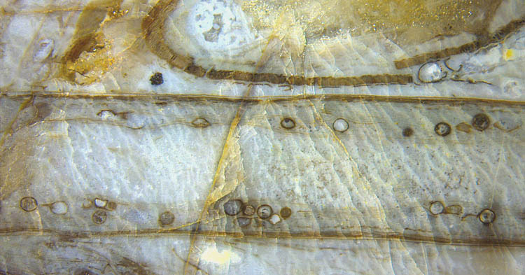
Fig.1: Lengthwise section
of Aglaophyton
with "acaulospores" of the
symbiotic fungus Glomites
rhyniensis; inclined section of empty
sporangium with broken palisade wall above. Image width 10mm.
The sample in Fig.1 represents a stage of decay
with no cortex tissue left and most spores having lost their
connection to the related vesicle which, when partially decayed and
looking like a tail, make what may be called a "tadpole
aspect". The empty
sporangium with
the characteristic palisade wall shows that the plant is really
Aglaophyton.
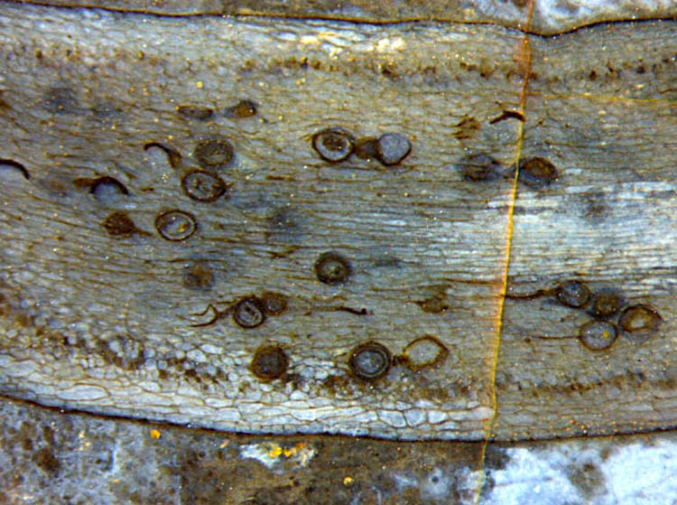 Fig.2:
Aglaophyton
with spore-saccule complexes of Glomites rhyniensis
inside exceptionally well preserved cortex tissue, also cells
filled with tiny
dark Glomites arbuscules seen
here in loose rows. Image width 5mm. (Note the magnification about
twice that of Fig.1)
Fig.2:
Aglaophyton
with spore-saccule complexes of Glomites rhyniensis
inside exceptionally well preserved cortex tissue, also cells
filled with tiny
dark Glomites arbuscules seen
here in loose rows. Image width 5mm. (Note the magnification about
twice that of Fig.1)
Additional information is
provided by Fig.2 with well preserved cortex tisse, dark dots of
Glomites arbuscules within cortex cells in two rows
at some
distance from the outer boundaries, and impressive examples of the
globular acaulospores, some of them seen with attached vesicle (Fig.3)
called saccule in [1].
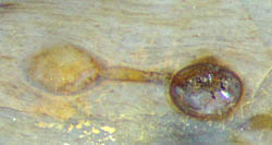 Fig3: Spore-vesicle complex of
Glomites
rhyniensis
Spore-saccule
complexes, separate spores, separate collapsed saccules, hypha
fragments, and two rows of dark dots, all
together in one image as in Fig.2, may be confusing but nevertheless
indicating Glomites.
Even
with less convincing fossil evidence as in Rhynie Chert News 182,
the collapsed
or cell-size
dark dots in shaky rows
are indicative of Glomites.
The
peculiar features shown here, as acaulospores grown only in connection
with vesicles of about same size, are not restricted to Glomites, as
pointed out in [4]. The present state of research concerning "the
abundance and morphological diversity of arbuscular mycorrhizal fungi"
has been characterized concisely as "... there is still
considerable undocumented diversity
in the form of new specimens and
hitherto unknown or unrecognized features of mycorrhizal fungi in the
Rhynie chert" [4]. That may justify this contribution
although
it does not really offer news. The sizes of the acaulospores (often
called
only spores) in this contribution
agree with those in [1], thus being much bigger than those in [4].
Samples:
Fig.1: Rh6_17.5 (2002); Fig.2: Rh11_10.1 (2003),
Fig.3: Rh7_10.2 (2003)
H.-J.
Weiss 2022
[1]
N.
Dotzler, Ch. Walker, M. Krings, H. Kerp, T.N.
Taylor, R. Agerer:
Acaulosporoid glomeromycotan spores with a
germination
shield from ... Rhynie chert.
Mycol. Progress (2009) 8, 9-18.
[2]
T.N. Taylor et al.: Fossil arbuscular mycorrhizae
from the Early Devonian,
Mycologia 87(1995), 560-73.
[3]
T.N. Taylor et al.: Fossil Fungi. Elsevier Inc.
2015, p.122.
[4] C. J. Harper,
Ch. Walker
et al.:
Archaeosporites rhyniensis gen. et sp. nov. (Glomeromycota,
Archaeosporaceae) from the Lower Devonian Rhynie chert: ...
Annals of Botany 126 (2020): 915–928.
 |
 |
187 |