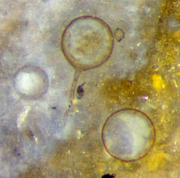
Conspicuous spheres in the Rhynie chert
Among
the often confusing multitude of decaying plant parts, spheres,
especially the larger ones, make conspicuous sights. They had been
hollow and filled with silica-rich water before subsequent
formation of silica gel and chalcedony.
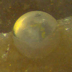
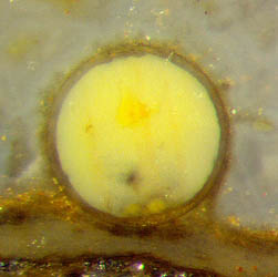
Figs.1-5: Conspicuous spheres in
Rhynie chert, possibly fungus
resting spores although no connection to
hyphae is seen here.
Frame sizes 0.6mm, sphere sizes 0.39,
0.33, 0.47, 0.44, 0.39mm.
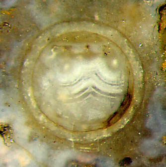
Fig.1 shows a solid
sphere half enclosed in chert. The formations in Figs.3,4 indicate that
there had been an intermediate stage with homogeneous silica
gel in the spheres which enabled spherulite
growth.
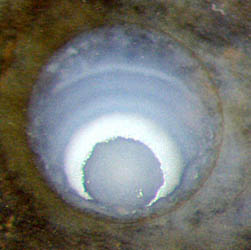
The walls in Figs.3,4,6 are seen in cross-section as very
thin dark circles. The
walls in Figs.5,7,8 might have been thick originally or become so later
by
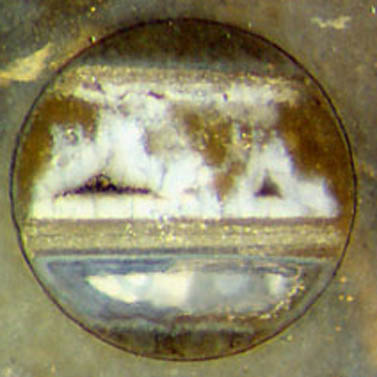
silica deposition. The big sphere in Fig.6 below seems to have been a
water tank before silicification, with floating microbial floccules
stained with iron oxides: Rhynie
Chert News 85.
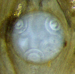
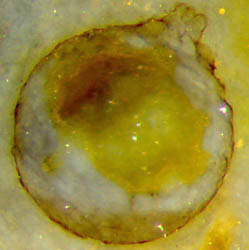
The occasionally
seen fungus hypha attached to a sphere as in Fig.9 seems to
justify the assumption that other spheres around, even in the absence
of hyphae, may also be fungus
resting
spores. Most
often this may be true but there are important exceptions. As a highly
wondrous fact, the cylindrical cells of the charophyte
green alga Palaeonitella
are
occasionally seen transformed into perfect spheres under
the influence of the parasitic fungi Milleromyces
rhyniensis and Krispiromyces discoides [1].
Hence,
in the absence of other
clues, one often cannot tell fungus resting spores apart from
transformed
alga
cells. The bomb-like
object in Fig.8 is certainly one of the latter. Spheres arranged in
pairs,
often interconnected,
are typical for bloated alga
cells, as in Fig.11. Nearby solitary spheres
(Fig.10) are most probably alga cells, too. As it is well
known
from other cavities in chert, agate-like fills can make fancy pictures.
Their complex structures can reveal details of the sequence of silicification
steps under changing conditions. One can say that several successive
deposition and dissolution processes had been at work in this sphere
once filled with silica-rich water, coupled to its surroundings by
diffusion.
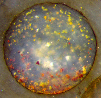
Figs. 6-9:
Spheres of different origin in
the Rhynie chert.
Frame sizes 0.8, 0.6mm. Sphere sizes 0.75, 0.6, 0.53, 0.17mm.
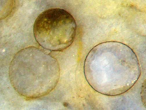 Figs.10,11: Spheres
paradoxically
formed from cylindrical cells of Palaeonitella
under the influence of parasitic fungi.
Figs.10,11: Spheres
paradoxically
formed from cylindrical cells of Palaeonitella
under the influence of parasitic fungi.
Frame heights 0.9mm. Left
sphere size 0.84mm.
All pictures: same scale, own finds.
For
the sake of simplicity and beauty, this contribution has been
restricted to larger spheres although smaller ones, too, may be highly
interesting, like the rare tiny capsules with an opening surrounded by
a collar, recently
discovered and ascribed to testate amoebae [2]. Similar
bomb-like capsules can be expected from broken dumbbell-like pairs
of spheres, as the one in
Fig.11. However, the
size difference
is large, and the similarity is incidental.
H.-J.
Weiss 2020
[1]
T.N. Taylor, M. Krings, E.L. Taylor: Fossil
Fungi. Elsevier 2015, p.64.
[2] Ch.
Strullu-Derrien, P. Kenrick, T. Goral, A.H. Knoll: Testate
Amoebae in the 407-Million-Year-Old Rhynie Chert. Current Biology 29
(Feb.2019), 461–467.
 |
 |
163 |