An uncommon fossil fungus
Fungus
hyphae and resting spores are abundant in the Lower Devonian Rhynie
chert. Often one can see dark cells arranged as a ring
on plant
cross-sections. The dark aspect is due to tangles of hyphae inside
cells, known as arbuscules, of the symbiotic glomeromycotan fungus Glomites rhyniensis
[1]. Other details of fungi in chert have been described in [1] and
elsewhere but many questions had to be left unanswered.
For example, the strange clusters of vesicles grown from the surface of
Aglaophyton
(Fig.1) are not mentioned in [1].
 The
arrangement of the clusters on the surface of Aglaophyton
resembles that of a blastocladian fungus in the Rhynie
chert
[1,2] but this similarity is most probably only superficial.
The
arrangement of the clusters on the surface of Aglaophyton
resembles that of a blastocladian fungus in the Rhynie
chert
[1,2] but this similarity is most probably only superficial.
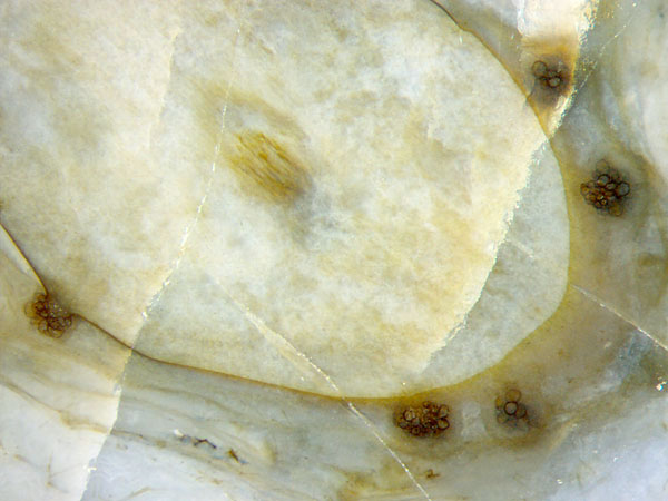
Fig.1: Clusters of fungus vesicles grown on the surface
of largely decayed Aglaophyton.
Width of the picture 4.3mm.
Except
for the poorly preserved central strand, no trace of plant tissue is
left in Fig.1. The contour of the section is marked by the cuticle.
When looked at edgewise, the cuticle appears
as a narrow black line, otherwise it is
seen as
transparent light-brown sheet, as in Fig.1 below right. As seen on the
left, and enlarged in Fig.2, the vesicles had grown on a mound bulging
out from the surface. Obviously the mound had been formed below the
cuticle, probably induced by the fungus in the
live plant.
The clusters on the right are not detached but thought to be
likewise connected to the slightly inclined shoot. The
sizes of the vesicles vary from 0.02mm (or smaller) to 0.1mm.
Their shapes may deviate from spherical by deformation due to mutual
contact.
Vesicles
of more or less globular shapes are known from numerous species of
fossil chytrids, a group of fungi thought to have separated from the
rest as early as during the Proterozoic [1]. The vesicles are
interpreted as
zoosporangia, with a "discharge pore" occasionally visible as a
characteristic feature [3]. Two big vesicles in Fig.2 have got a kind
of
short funnel, very dimly seen, which might have served as a
discharge pore. (Note the pale short extension pointing left on the big
vesicle on the left.)
Hence, the assumption may be justified that
the clustered vesicles shown here are zoosporangia of a fungus of the
chytrid clade. This implies the following statement: The vesicles
presented here are bigger than any fossil chytrid zoosporangium
described in [1].
The flower-like aspect of the cluster in Fig.3
(right) is an illusion: One sees only the bigger outer vesicles while
most of the cluster is unseen in the depth.
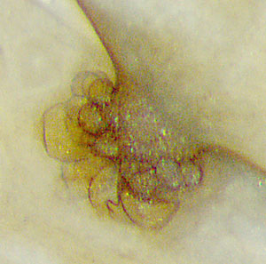
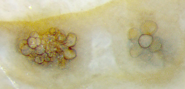
Figs.2,3: Details from Fig.1, clusters most probably of
chytrid zoosporangia. Note the bulge grown below
the cuticle (Fig.2) and the transparent light-brown cuticle in Fig.3.
Height of the images 0.5mm.
H.-J.
Weiss 2017
Annotation
Nov.2017:
According to M. Krings (private communication), this fungus is the one
which has been
described in [4] as Trewinomyces
annulifer. That one is really
similar to this one but there are differences. The sporangia in
[4] are mostly pear-shaped or elongate and sit on
stalks while these sporangia are mostly globular, and no stalks are
seen in these figures. The sporangia diameters
in [4] are smaller, up to 65µm, compared to 115µm here.
According
to [4], "Assemblages of zoospore fungi usually consist of individuals
at different developmental stages" but "the growth of T. a.
in tufts in which all individuals appear to be at approximately the
same stage of development" is thought to be an argument against an
affinity to the Chytridiomycota and Blastocladiomycota. The quotations
rather favour such affinity of this fungus: As seen in these images,
the assemblages really "consist of individuals
at different developmental stages."
So we are left with two
alternatives: Either the genus diagnosis of T. a. has to be
emended such that it includes this fungus, or this fungus is another
species.
[1] T.N.Taylor, M. Krings, E.L. Taylor:
Fossil Fungi, Elsevier 2015.
[2] W. Remy, T.N.
Taylor, H. Hass: Early Devonian fungi. Am. J.
Bot. 81(1994),
690-702.
[3]
Ch. Strullu-Derrien, T. Goral, J.E. Longcore, J. Olesen, P. Kenrick,
G.D. Edgecombe:
A New Chytridiomycete Fungus Intermixed with Crustacean Resting Eggs
in
a 407-Million-Year-Old Continental Freshwater Environment.
PLoS ONE
11(12): e0167301. doi:10.1371/journal (2016).
[4] M. Krings, T.N. Taylor, H.
Martin:
An enigmatic fossil fungus ... , Mycologia 108 (2016), 303-312.
 |
 |
108 |