Microbial reefs in the Devonian
swamps at Rhynie

Microbes are abundantly present in the Rhynie chert but
often not recognized as such. They may appear as mm- to cm-size
bulging clouds, apparently structureless, and
may remain unnoticed unless
more conspicuous, as those in Rhynie
Chert News 68.
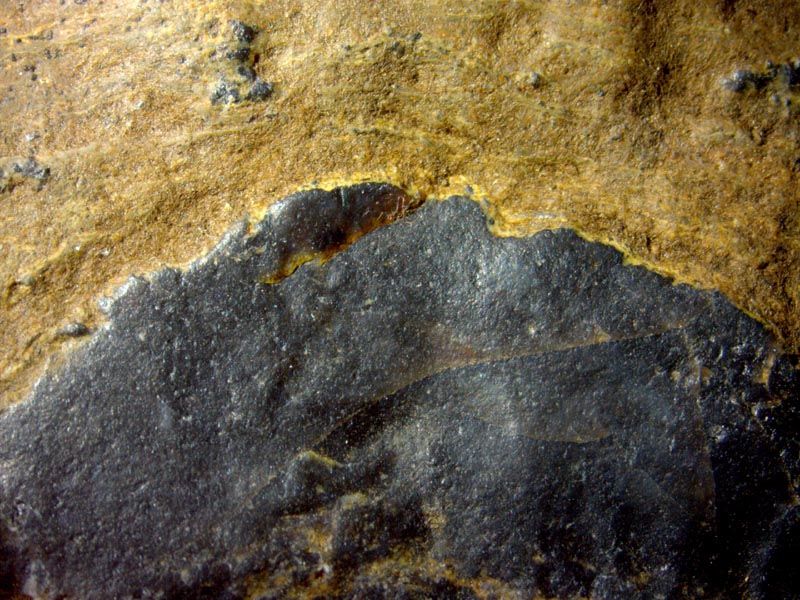
In the present sample there are microbial clouds or blobs clearly
separated from their yellowish surroundings, as
in Fig.1.
Fig.1: Raw outside of a sample from Rhynie with bulging top of a chert
layer and yellow sediment above. Width of the image 17mm.
The bulging cloud in Fig.1 at
the top of the chert layer, like everything else now seen in the chert,
had grown or formed in water. Judging from the distinct boundary, the
cloud had not been a loose assembly of microbes when the yellow
sediment settled. There is no indication of deformation under load,
hence one can assume that the dark cloud had been solid in the swamp
water before. This is not surprising since differential rates of
silicification of objects in the swamp is a well-known fact.
Possibly
the microbes kept themselves together in a big cloud or blob by some
kind of organic gel between them, which triggered early silicification
while the surrounding water remained fluid. Apparently a subseqent
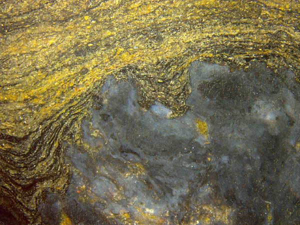
muddy flow did not affect the craggy shapes, and the mineral debris
had not entered but accumulated above (Figs.1-3), which invokes the
idea of buried reefs.
The nature of some details seen here and occasionally with similar
microbial formations in other samples, as irregular
black inclusions of various size and
shape as in Fig.2, remains obscure.
Possibly
the microbial
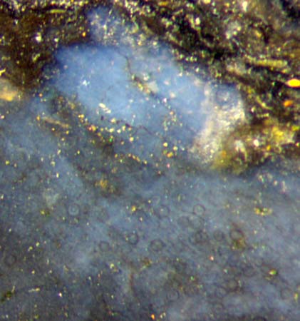
clouds could have offered suitable places for other organisms to
thrive: An assembly of transparent spheres is seen in
one cloud of this sample: Fig.3. (Annotation and deletion Nov. 2017:) Apparently they belong to the
same fungus species as shown in Rhynie
Chert News 115 and interpreted there as Zwergimyces.
Fig.2 (left): Microbial formations like
reefs: not deformed by the silt deposited later.
Cut face, same sample as
Fig.1 but other position. Width of
the image 11mm.


Fig.3: Middle of Fig.2 enlarged such that transparent spheres of sizes up to about 50Ám become
visible.
Width of
the image 1.3mm.
H.-J.
Weiss
2017
 |
 |
113 |


