Early land plant aging
 Aglaophyton
is the most common fossil
plant in the Lower Devonian Rhynie chert but in rare cases like this
one it offers surprising
aspects which may give rise to contemplations concerning details not
thoroughly considered before. The most conspicuous detail in this image
is the white ring around the
central xylem. It simply
indicates that, for whichever reason, the chalcedony had recrystallized
there into silica grains
of such size that they
reflect the incident light like snow. (A similar ring is seen
in Rhynie
Chert News 70.
As
a very rare phenomenon, tiny white dots around the xylem are seen
in Rhynie
Chert News 2.)
It is not known whether or not this white ring can provide relevant
information on silicification processes.
Aglaophyton
is the most common fossil
plant in the Lower Devonian Rhynie chert but in rare cases like this
one it offers surprising
aspects which may give rise to contemplations concerning details not
thoroughly considered before. The most conspicuous detail in this image
is the white ring around the
central xylem. It simply
indicates that, for whichever reason, the chalcedony had recrystallized
there into silica grains
of such size that they
reflect the incident light like snow. (A similar ring is seen
in Rhynie
Chert News 70.
As
a very rare phenomenon, tiny white dots around the xylem are seen
in Rhynie
Chert News 2.)
It is not known whether or not this white ring can provide relevant
information on silicification processes.
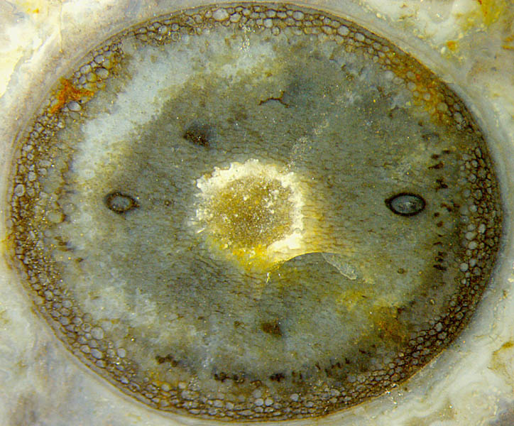
Fig.: Cross-section of Aglaophyton
with several competing
phenomena of selective
decay and preservation, recorded in progress by silicification. Size
5mm.
The
ring of well-preserved tissue consisting of distinctly visible cells
below the poorly visible epidermis is doubtless related to
silicification in a non-trivial way. The preservation of the tissue up
to a certain depth is not simply due to a finite penetration depth of
dissolved silica into the
dead plant by diffusion
as claimed in [1] but had been governed by the live plant instead, as
emphasized in Rhynie
Chert News 181.
The present cross-section is particularly interesting as it shows an
intermediate stage on the way towards a final "hollow
straw"
stage. The peripheral layer of well-preserved cortex
cells is that part of the tissue which had been made decay-resistant.
It has persisted
while other
areas of the cortex tissue are seen in a more or less decayed state.
Black spots, loosely arranged at a distance from the epidermis, are
cortex
cells filled with a tangle of
tiny hyphae (arbuscules) of the
symbiotic
fungus Glomites
rhyniensis
[2,3], partially
collapsed.
Obviously this cross-section offers a
rare sight on details
while most other sections
of Aglaophyton,
though abundantly seen on
cut faces or fracture faces of the chert layer, do less
so. The
two thick-walled black capsules are
probably "acaulosporoid
glomeromycotan spores" [3] (resting spores) of
Glomites. Their symmetrical
arrangement
may be misleading: It is purely incidental, as can be guessed from Rhynie
Chert News 55, 60, 97, 142, 187.
Sample:
Rh15/81.1 (0.32kg) obtained
from Barron
in
2014.
H.-J.
Weiss 2021 revised version 2022
[1] www.abdn.ac.uk/rhynie, Chapter Taphonomy.
[2]
T.N. Taylor et al.: Fossil
arbuscular mycorrhizae from the Early Devonian, Mycologia
87(1995), 560-73.
[3]
T.N. Taylor et al.: Fossil Fungi. Elsevier Inc.
2015, p.122.
 |
 |
182 |