A rare aspect of Aglaophyton
Aglaophyton major,
formerly known as Rhynia
major [1], is one of the
plants found in the Rhynie chert whose position in the phylogenetic
tree is being disputed. The absence of typical tracheids in the
conducting strand led D.S.
Edwards [2], after comparison with
the anatomy of mosses, to the conclusion that this plant is not a
vascular plant and therefore not related to Rhynia gwynne-vaughanii,
which he regarded as a sufficiently strong reason to change its name.
There remains the intriguing fact that, apart from the tracheid wall
pattern, the two plants are more similar to each other than to any
other Rhynie chert plant discovered hitherto. Particularly obvious is
the
similarity of their sporangia: In either species the sporangium wall
consists of three layers of tissue. The outer one is easily recognized
on sections by its distinct palisade-like array of cells arranged such
that the sporangium appears twisted in
side view. (The twist is not brought about by lengthwise splitting of
the sporangium as proposed
in [2] as it is clearly seen on the non-split sporangium.)
These and other similarities nurture the suspicion that the two plants
may not be as unrelated as suggested by the absence of tracheid wall
thickenings in Aglaophyton
[3]. So it will be interesting to look for
more evidence supporting one or the other view.
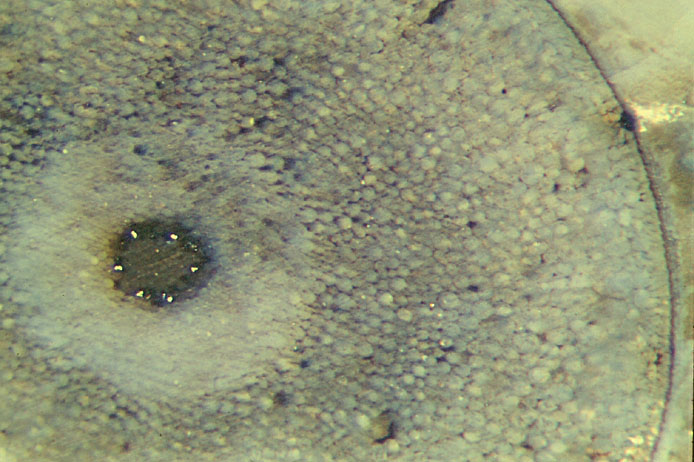
Photograph: Aglaophyton
cross section with unique "fairy ring" resembling the array of
protoxylem in the xylem strand of a vascular plant. Note also
the incidentally cut stoma on the right.
Sample:
Rh2/73, 0.29kg, Part 1, obtained from J. Shanks in 2002.
Unexpectedly, one small sample of chert with uncommonly well preserved
Aglaophyton axes
has provided a rare sight as shown here: The
"fairy ring" of distinct white spots can hardly be regarded as
incidental although it is seen in this regularity on only one section.
On other sections, the spots, if there are any, do not form a complete
ring or are more irregularly distributed. As a possible explanation,
some slight chemical difference between cell types, inherent or due to
differential stages of decay, led to different mineralisation resulting
in strong optical contrast. (Slightly differing starting conditions
leading to vastly differing final stages via complex processes is not
uncommon.) The white spots are brought about by strong
reflection, probably due to fine-grained quartz, contrasting to the
more or less transparent chalcedony.
In the particular case seen in Fig.1 the situation seems to have been
so subtly balanced that only a certain type of cells has been affected
by the contrast-enhancing process. Comparison with the well-known
arrangement of tissues in other simple vascular plants suggests that
this central strand is made up of xylem with 6 spots of protoxylem.
This interpretation may provide additional evidence in favour of the
assumption that Aglaophyton
does not as much differ from a vascular
plant as assumed in [2] but is simply an unusual vascular plant without
the usual tracheid wall pattern.
H.-J. Weiss
2005
[1] R.
Kidston, W.H. Lang: On Old Red Sandstone plants showing
structure from the Rhynie Chert
bed, Part II,
Trans. Roy. Soc.
Edinburgh 52(1920), 643-80.
[2] David
S. Edwards: Aglaophyton
major, a non-vascular
land-plant from the Devonian Rhynie Chert,
Bot. J. Linn. Soc. 93(1986), 173-204.
[3] Dianne
Edwards : A review of the sporophytes of embryophytes
in the cherts at Rhynie,
Trans. Roy. Soc. Edinburgh, Earth
Sciences 94(2004 for 2003), 397-410.
 |
 |
2 |