Enigmatic hollow straws of early land
plants
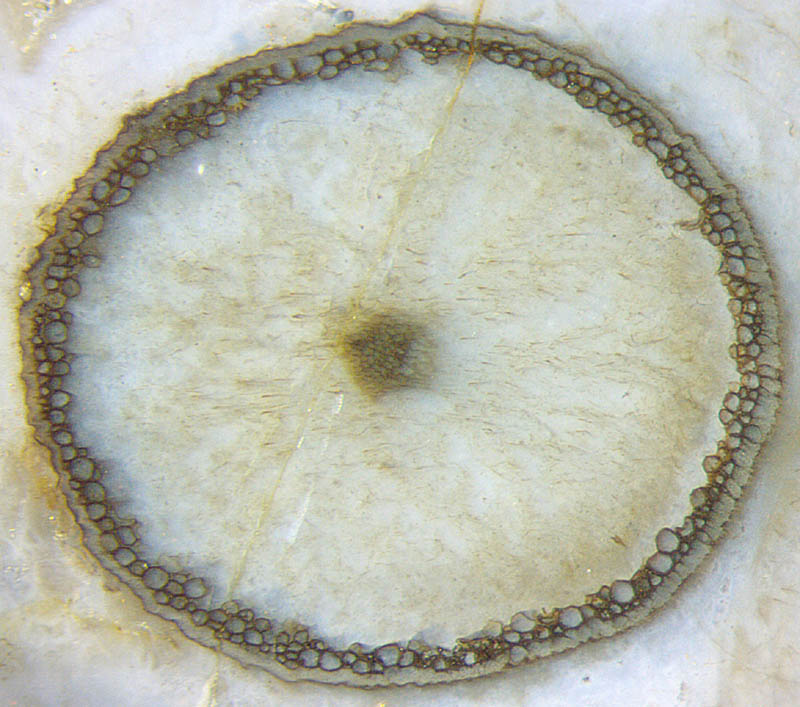 Hollow straws of early land plants are occasionally
seen on sections of Rhynie chert as circular rings with well
preserved cells while most of the tissue is missing (Fig.1). An
explanation as
a result of limited diffusion depth of silica in the dead plant lying
in the swamp water, as proposed in [1,2],
is rejected here. Not the epidermis as the outermost layer but the
tissue immediately below is well preserved, usually with cell walls
stained dark.
Hollow straws of early land plants are occasionally
seen on sections of Rhynie chert as circular rings with well
preserved cells while most of the tissue is missing (Fig.1). An
explanation as
a result of limited diffusion depth of silica in the dead plant lying
in the swamp water, as proposed in [1,2],
is rejected here. Not the epidermis as the outermost layer but the
tissue immediately below is well preserved, usually with cell walls
stained dark.
Fig.1: Cortex tissue well-preserved immediately below the
epidermis but decayed and vanished elsewhere. Width 2.5mm.
This could
not have been brought about by mere diffusion. Apparently the living
plant had prepared, by unknown means, part of the tissue so that it
served as a protection against some intruder, with
the side effect that the tissue thus prepared persisted
while the majority of the tissue decayed and vanished before becoming
silicified.
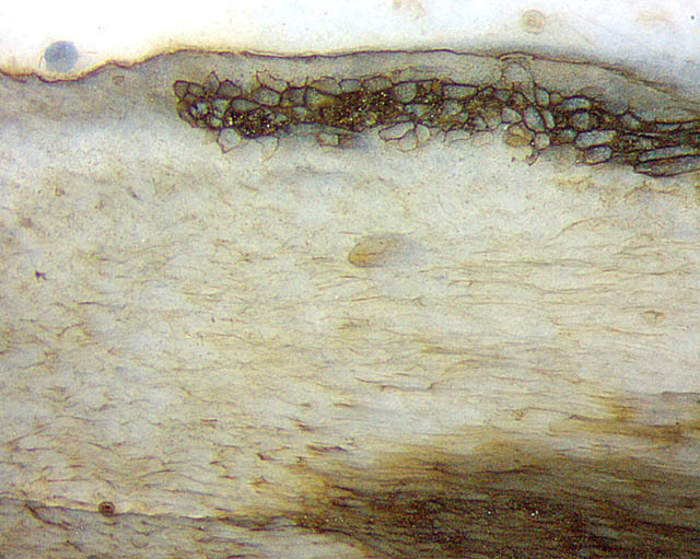
Such an interpretation involving something more complex than mere
diffusion
could be compatible with the sudden onset (or ending) of the
strip of black-walled cells seen on the inclined cut in Fig.2. The
central strand had not been affected since it has retained its
light-brown stain. This applies also to the poorly visible cells on the
inclined cut face of another sample in Fig.3.
Fig.2
(below): Sudden onset (or ending) of a strip of well-preserved cortex
cells below the epidermis.
Image width 2mm, same scale as
above.
The cut face of the chert in Fig.3 unexpectedly reveals a rare
phenomenon providing information concerning the processes
involved here. A hole in the layer of black-walled cells inside the
hollow straw is now covered with a dome-shaped layer of cells of the
same type.
This dome could not have grown in the empty straw. It must represent, like
the black layer of tissue below the epidermis, the remaining cortex
spared from decay after it had been made decay-resistant by the live
plant. (The cortex tissue had formerly filled the whole space between
epidermis and central
strand.) Undoubtedly
the transformation of cortex tissue into the persistent dome covering
the damaged
spot had been governed by the plant with the aim to
prevent further damage. The combination
of persistent dark dome and persistent dark peripheral
layer suggests that the latter, too, had been prepared by the live
plant to
keep out intruders.
Fig.3 (below): Protective dome formed from cortex cells formerly present
throughout before part of the cortex cells was made persistent and
became black-coated
while the others decayed and vanished, thus leaving a hollow straw.
Image width 10mm.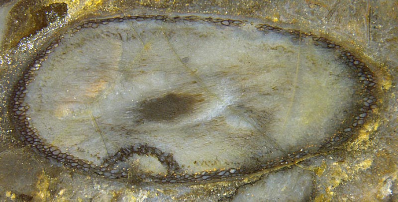
As
a side effect, the persistence of the tissue favoured the formation of
the black coatings before all turned into silica gel and chert.
The
interpretation of these hollow straws as Aglaophyton seems
justified by the observation that typical details of this plant, as the
"palisade wall" of its sporangia, can be found nearby.
Samples: Rh12/91.3+5 (2006) Figs.1+2;
Rh12/162.2 (2007) Fig.3.
H.-J.
Weiss 2021
[1] C.L.
Powell, N.H. Trewin, D. Edwards: Palaeoecology and plant
succession
in a borehole through the Rhynie cherts, ...
Geological Society, London,
Special Publications 180 (2000), 439-457.
[2] www.abdn.ac.uk/rhynie, Chapter Taphonomy.
 |
 |
181 |