Selective decay resistance of soft
tissue in early land plants (1)
To be more specific, this is about the
conspicuously high decay resistance of small fractions of cortex tissue
often but not always found in Aglaophyton
and Ventarura
[1], the most common and the
rarest land plant preserved in the Early Devonian Rhynie chert. For
an introduction it is mentioned here
that the xylem of the central strand is a comparatively "hard"
tissue, judging from its narrow cells and the fact that it is usually
seen in
good shape even when other parts of the plant are in a queezed or
decayed state. Most of
the cross-section, which is the area between the
epidermis and the phloem around the
xylem, is made up of a tissue
called cortex .
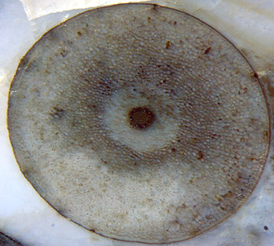
Fig.1 (right): Aglaophyton
cross-section, 4mm, with dark xylem, concentric lighter phloem,
epidermis, and cortex as the largest part in between.
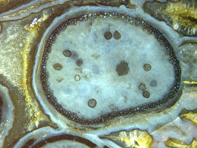 Fig.2
(left): Aglaophyton
as a hollow straw, with xylem strand and fungus vesicles inside, early
silica gel and microbial coatings
outside, and a ring of well-preserved
cells.
Fig.2
(left): Aglaophyton
as a hollow straw, with xylem strand and fungus vesicles inside, early
silica gel and microbial coatings
outside, and a ring of well-preserved
cells.
The cortex serves more than one
purpose: providing a place for photosynthesis, stuffing the tube
and keeping it in shape, and fixing the central strand in the centre.
All this can be achieved with a smaller amount of cortex, judging from
live plants with large voids
in the cortex. Possibly the
plant can do with even less cortex, as a (partially) hollow straw.
Hollow straws of Aglaophyton,
seen in cross-section as a ring of surprisingly well preserved
cells, are not rare (Figs.2-4).
The
existence of a cylindrical lining involving well-preserved
cortex cells while the bulk of the cortex has completely vanished
gave rise to wonder.
The proposed explanation as a result of quick silicification of only a
few peripheral layers of cells [2,3] by moving "silicification fronts"
has been doubted for various reasons (as by A. Channing [4]).
It seems to be
disproved by Fig.3, where Aglaophyton
sections with
different aspect are seen close together and hence had been subjected
to the same silicification conditions.
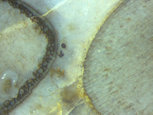 Fig.3 (left): Aglaophyton
sections with strongly differing aspect.
Fig.3 (left): Aglaophyton
sections with strongly differing aspect.
Obviously something else than silicification
fronts is required here for an explanation. The possibly
complex processes producing the phenomenon may formally be reduced to
something simple: It was not silicification but decay resistance of the
peripheral layers which came first.
It would be interesting to know whether or not the plant went on
living as a hollow straw for a while with the
bulk of the cortex being more or less destroyed. 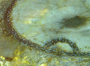 A
particular search for related evidence has not yet been done.
A
particular search for related evidence has not yet been done.
Fig.4 (right): Aglaophyton,
cortex decayed except for a layer of well-preserved cells along the periphery, with a dome-shaped extension of the decay-resistant layer
covering a spot of
damage. Width of the dome 2mm.
There
is evidence that the persistent tube is not brought about simply by
providing some
anti-rot
agent to a peripheral layer of cells. Fig.4 shows
that a more complex process
must have been involved: It is seen that
the plant
has managed to form a dome-shaped decay-resistant
cover above a spot of local
damage.
What makes the sections of hollow straws of
Aglaophyton
(as in Figs.2-4) still more conspicuous is a black stain or deposit
often but not always seen on the cell
walls, which makes the straws stand out starkly against the pale plant
debris in the chert. So it appears that in addition to a
decay-resistant lining consisting
of tissue, there is a lining on smaller scale, consisting of a deposit
on
decay-resistant cell walls.
The
phenomenon of highly selective preservation amidst general decay has
become even more enigmatic with the discovery of Ventarura, whose
cross-sections are often but not always conspicuous for a concentric
ring of similar aspect as the rings in Figs.2-4 but with the
distinction that it is placed somewhere within the cortex, most
often well away from the (usually decayed) epidermis (Fig.5).
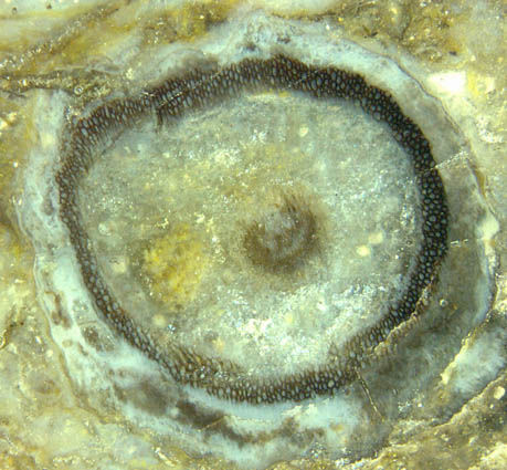
Fig.5: Ventarura
cross-section, 7mm, with a ring consisting of
well-preserved
cortex cells while the epidermis and the cortex within and without the
ring are completely decayed.
Similar as with Aglaophyton,
good preservation and dark aspect are not coupled here. If not dark,
the well-preserved fraction of the cortex is easily overlooked, in
which case the plant fragment may not be recognized as Ventarura
at first sight. Cell walls with and
without black stain can be present in the same ring and
even in the same cell, as seen in Fig.6.
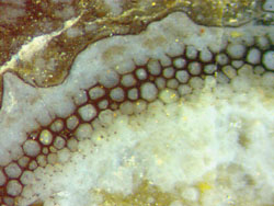
Fig.6 (right): Ventarura
cross-section, detail. Note the cells with or without dark lining, also
both with and without dark lining
on parts of the cell wall, and walls which do
not seem to make a closed cell anymore.
Apparently the claim in [1] that
the cells with thick-walled aspect are sclerenchymatic can
be refuted by the evidence presented here. This is also
confirmed by
Fig.7 where it is seen that the black lining can break off in flakes
and hence
most probably is not an intrinsic component of the cell wall but a
deposit. Where the ring had lost its dark aspect by such subsequent
process whose nature is likewise unknown as that of the deposition,
the original thin cell walls have reappeared, well
seen with suitable illumination but less so in Fig.7,
with a few black splinters of the deposit still sticking to them.
By the way, Fig.7 also shows that Ventarura
can
easily be mistaken for Aglaophyton
if the rot-resistant layer is found close to the
outer boundary after the cortex has decayed and
vanished there. In this connection it may be mentioned that, the other
way round, Aglaophyton
occasionally had been mistaken for Ventarura.
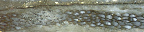
Fig.7 (right): Ventarura,
detail of inclined section, well-preserved ring of cells with dark
deposit on the walls, flaked off locally.
Summary
(1) Highly decay-resistant cortex areas, ring-shaped
on cross-sections, are seen often
at
the periphery of Aglaophyton and
regularly in Ventarura.
(2)
The decay-resistant tissue can favour or induce a deposition process
in the dead plant, making the cell walls appear dark and strong.
(3) The tissue with coated cell walls in Ventarura has been
mistaken for sclerenchyma.
(4) The similar aspect of the persistent tissue in the two non-related
plants
suggests similar formation, which may help to explain either.
(5) Providing decay resistance and stain to
some cortex fraction may not always affect cells as a whole
but often only part of the wall.
(6) The question remains which kind of processes could possibly produce
such enigmatic outcome.
H.-J.
Weiss
2014
slightly revised 2015
[1] C.L.
Powell, D.
Edwards, N.H. Trewin: A new vascular plant from the
Lower Devonian Windyfield chert, Rhynie, NE Scotland.
Trans. Roy. Soc. Edinburgh, Earth Sci.
90(2000 for 1999), 331-349.
[2] C.L.
Powell, N.H. Trewin, D. Edwards: Palaeoecology and plant
succession in a borehole
through the Rhynie cherts, ...
Geological Society, London,
Special Publications 180 (2000), 439-457.
[3] www.abdn.ac.uk/rhynie, Chapter Taphonomy.
[4] A. Channing:
Processes and
Environments of Vascular Plant Silicification: Thesis, Chapter
6, Cardiff University, 2001.
 |
 |
60 |