Palaeozoic wood rot mistaken for
oribatid mite coprolites
Conspicuous tiny clots related to local damage in the wood of fossil
trees are usually interpreted as coprolites, also more specifically as
mite coprolites or even oribatid mite coprolites. Recently, evidence
contradicting such interpretation has been compiled: see
Misconceptions,
Oribatid mite coprolites. It
is worth mentioning that most evidence is taken from published pictures
whose authors did not notice essential details. As the proponents of
the coprolite hypothesis are reluctant to accept contrary arguments,
more evidence is extracted from their publications here.
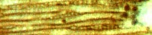 Fig.1:
Lower Permian wood from Schallodenbach, Germany.
Fig.1:
Lower Permian wood from Schallodenbach, Germany.
Note the small clot deep inside a tracheid. Detail of Fig.30
in [1].
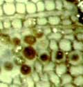
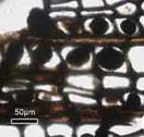 Fig.2 (left): Lower Permian
wood, Schallodenbach; more of it here.
Fig.2 (left): Lower Permian
wood, Schallodenbach; more of it here.
Fig.3: Lower Permian wood, detail of Fig.4F in [2].
The clots are essentially globular when smaller than the cell
cross-section.
The arrangement of the clots hints at a process of clot formation
propagating from cell to cell.
Compared to the tube-like xylem cells, the clots appear globular. They
may be really globular as long as they are smaller than the cell
cross-section, as in Figs.1,2,3. Bigger clots either fit nicely into
the angular cell lumen and keep their shape after the cell walls have
gone, as in Figs.4,5, or they expand and deform the cell and remain
essentially globular. It is not known under which conditions one or the
other of these options, or some intermediate stage, is realized.
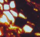
 Fig.4
(left): Psaronius aerial root cross-section, phloem cells empty or
completely filled with clot matter. Detail of Fig.8 in [3].
Fig.4
(left): Psaronius aerial root cross-section, phloem cells empty or
completely filled with clot matter. Detail of Fig.8 in [3].
Fig.5: Lower Permian wood, angular clots released after disintegration
of tissue.
Detail of Fig.3I in [2].
The observations suggest that the clots grew inside the cells, and are
no mite droppings fallen into the open end of damaged tube-like cells
and slid down as claimed by the supporters of the
coprolite idea [4]. Sliding down a tube under its own weight is a
notion which does not apply to things as tiny as cell-size clots
because adhesion forces would exceed their
weight and make them stick to the wall, all the
more so in the presence of traces of
moisture. Obviously the clots seen inside the tubes in Figs.6,7,8 grew
there on the very spot where they are now, judging from the bulges.
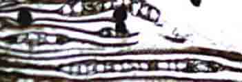
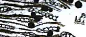
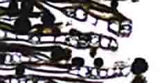
Figs.6,7,8: Permian wood with clots inside the tube-like tracheids
causing them to bulge, which indicates that the clots grew there and
hence are no coprolites. Details of Fig.4D in [2].
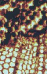 Fig.9:
Lower Permian wood, clots interpreted as coprolites in [3] although
they are arranged in rows, slightly tilted to the right but compatible
with the files of empty cells below. Detail of Fig.17
in [3].
Fig.9:
Lower Permian wood, clots interpreted as coprolites in [3] although
they are arranged in rows, slightly tilted to the right but compatible
with the files of empty cells below. Detail of Fig.17
in [3].
The rows of clots in Figs.2,3,9 suggest a formation process spreading
along cell files. This is also suggested by pith rays filled with dark
substance in Fig.10 and in [3], Fig.17, and by observations on [2],
commented in [5]. 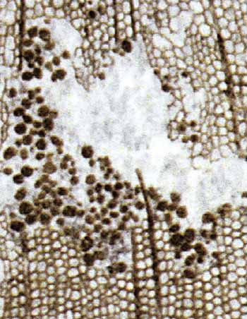 The
tissue in the upper half of Fig.9 is deformed but apparently still
coherent. Hence, what is described as a frass gallery in [3],
Fig.17, is locally deformed wood affected by a process producing dark
substance inside the cells. The narrow vertical line crossing some
clots is a crack formed at an early stage of silicification.
The
tissue in the upper half of Fig.9 is deformed but apparently still
coherent. Hence, what is described as a frass gallery in [3],
Fig.17, is locally deformed wood affected by a process producing dark
substance inside the cells. The narrow vertical line crossing some
clots is a crack formed at an early stage of silicification.
Fig.10 (right): Lower Permian wood cross-section with alleged "mite
borings and galleries containing coprolites", detail of Fig.6C in [2],
interpreted here as wood rot involving the formation of dark substance
in the cells and pith rays, with subsequent disintegration of the
tissue.
As a conspicuous feature, the clot sizes vary strongly, and so do the
cell sizes in this
particular sample. Some clots may have grown bigger than the tracheid
diameter, as in Figs.6,7,8.
A large part of the clots are angular, as distinctly seen above left.
The angular clots preserve the shape and size of cell cross-sections
after the cell walls have dissolved, as emphasized in [6].
A few clots are still inside cells in areas where the affected tissue
is not yet
disintegrated.
The process producing the dark substance has entered into and spread
along part of the pith rays, delaying their disintegration. One
affected pith ray is seen preserved where the surrounding tissue is
not. More pith rays with dark fill are seen in [2], Fig.6C, and in [3],
Fig.17. The dark area in Fig.3 is possibly also a filled pith ray.
To summarize, all clots seen in these pictures, except for Fig.2, have
been mistaken for coprolites in the scientific literature. Fig.2 is
taken from an own sample found and kindly provided by Ch. Krüger,
Schallodenbach. The arrangement of the clots in Fig.2 raised doubts
concerning the coprolite interpretation. The doubts increased with
every careful inspection of publications on the subject. A set of
simple rules has been proposed in [7] as an aid for telling apart true
and false coprolites.
The question remains what the clots are if not coprolites. A similarity
to clusters of very thin hyphae of the fungus Glomites rhyniensis
in cells of Lower Devonian plants is pointed out in [8]. However, Glomites is known
to propagate through the intercellular space with coarse hyphae in
addition to the thin ones inside cells but no coarse hyphae have been
seen in the tissues with the clots considered here. So one has to
consider formations by other fungi or by microbes as
potential explanations for the clots.
H.-J. Weiss
2011
[1] R.
Noll, V. Wilde : Conifers from the „Uplands“
– Petrified wood from Central Germany,
in:
U. Dernbach, W.D. Tidwell : Secrets of Petrified Plants,
D'ORO
Publ., 2002. 88-103
[2] Zhuo
Feng,
Jun Wang, Lu-Yun Liu :
First report of oribatid mite (arthropod) borings and coprolites in
Permian woods from the Helan Mountains of
northern China.
Palaeogeography,
Palaeoclimatology, Palaeoecology 288(2010), 54-61.
[3] M.
Barthel, M. Krings, R. Rößler: Die
schwarzen Psaronien
von Manebach, ihre Epiphyten, Parasiten
und Pilze.
Semana* 25(2010),
41-60. * recently re-named, former
name: Veröff. Naturhist. Mus. Schleusingen.
[4] Z.
Feng, R. Kretzschmar: private communication.
[5] Dubious oribatid mite coprolites once more: Comment on
Z.
Feng et al.
(2010),
[6] Alleged coprolites - Remnants of decayed
tissue.
[7] Alleged arthropod coprolites re-interpreted.
[8] Alleged coprolites
of "unknown creatures" replace alleged oribatid mite coprolites.
Annotation: A critical comment
on [3] (in German) suitable for publication in the journal "Semana" has
been rejected on grounds of pretended copyright violation.
|

|
 8 8 |