Dubious oribatid mite coprolites once more: Comment on Z.
Feng
et al. (2010)
Lately, not much was heard about oribatid mite coprolites, a favourite
topic in palaeobotany a few years ago. (See chapter
Misconceptions.)
So the recent paper by Feng
on this subject [1] came
unexpected. Unfortunately, it does not fulfill the expectations raised
by its title.
A brief look at the photographs of dark clots in silicified wood
reveals that most of them are no coprolites. Although the subject had
been discussed in detail in connection with previous papers on alleged
mite coprolites so that one might expect the matter settled, it has to
be considered here anew, with the same old and simple but adequate
reasoning applied to the new pictures provided in [1].
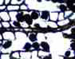
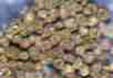
There are several conspicuous details in the pictures which should
raise suspicion even with unexperienced observers. One such detail is
the presence of clots with distinctly angular shape (Figs.1...4).
Furthermore, the shapes and sizes of the clots often agree with the
shapes and sizes of nearby cells, as observed before on fossil wood
from Antarctica.
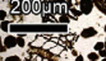
Figs. 1...4: Clots in silicified Permian wood, interpreted
as mite coprolites in [1] despite of their angular shapes; details from
[1], there in Figs. 3I, 4E, 3C, 3I (left to right).
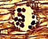
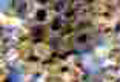
Among the numerous clots in the samples there are often
a few ones in positions which seem awkward from a coprolite point of
view: They are seen deep inside tracheids where no mite could have
crept in. The deceptively plausible explanation offered by the
proponents of the coprolite hypothesis that they simply fell into the
tracheid at an open end is not a good one: In some cases the clots are
seen in several neighbouring tracheid cross-sections (Figs. 5...7), so
while falling in they all would have got stuck at the same depth. By
the way, things as small as mite droppings would not easily tumble down
tubes of about the same diameter since their weight is usually
negligible compared to adhesion forces, especially if traces of
moisture are present. They would just stick to the wall but not slide
down.
The arrangement of the clots in the below pictures suggests quite
another explanation: They have been formed inside the cells. The two
tiny clots in neighbouring cells in Fig.6 are possibly early stages of
clot formation. As a significant feature also observed with samples
from elsewhere, the clots are not randomly
distributed among the cells but tend to cluster.
In other words, a cell
with clot is more likely to have got a neighbouring cell with clot than
to have not. This supports the previously proposed idea of a fungus
infection
spreading from one cell to another as the cause of clot formation
[2,3]. A phenomenon of this kind, a clot sending a hypha through the
cell wall to make another clot in the neighbouring cell, is seen in a
remarkable photograph in [4].
Some of the clots eventually keep their position even after the cell
walls have decayed, as three clots in a row in Fig.5, and less clearly
seen in Fig.6. The most convincing evidence for clot formation in cells
is provided by Fig.7, with three and four clots in a row, sitting at
the same height in neighbouring tracheids. Note also the two smaller
clots in smaller cells below, one above the scale bar and another one
on the right. Some of the clots look as if their shapes were tapering
towards a site of attachment to the cell wall.
 Figs.
5...7: Clots in silicified Permian wood, interpreted as mite coprolites
in [1] despite of their peculiar arrangement suggesting their formation
inside cells; details from [1], there in Figs. 4B, 4B, 4F (left to
right).
Figs.
5...7: Clots in silicified Permian wood, interpreted as mite coprolites
in [1] despite of their peculiar arrangement suggesting their formation
inside cells; details from [1], there in Figs. 4B, 4B, 4F (left to
right).
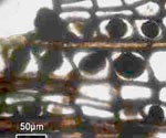
A
formation in silicified wood with not really clot-like aspect
(Figs.8...10) is also interpreted as oribatid mite coprolites in [1].
There are patches on the petrified wood cross-section where the usual
wood structure with cells in radial files (vertical in Figs.8 and 10)
is replaced by something resembling an imperfect honeycomb structure.
 Figs.
8...10: Silicified Permian wood affected by what appears to be some
kind of wood rot, interpreted as mite coprolites in [1] despite of the
polygonal shape of the elements and their dense-packed honeycomb-like
arrangement; details from [1], there in Fig. 6E.
Figs.
8...10: Silicified Permian wood affected by what appears to be some
kind of wood rot, interpreted as mite coprolites in [1] despite of the
polygonal shape of the elements and their dense-packed honeycomb-like
arrangement; details from [1], there in Fig. 6E.
As seen in
Figs. 8 and 10, the light-coloured area adjoins the unaffected wood
whose cells appear dark as they were hollow before silicification and
are now filled with clear chalcedony so that one can look into the dark
depth. The two light-coloured elements below the empty cells in Fig.10
fit into the cell pattern seen above. So they must be cells filled with
some substance.
In order to interpret the alleged coprolites in
Figs. 8 and 9 as modified cells, it must be explained why they are not
arranged in files and seem to be slightly larger than the cells. As a
possible explanation, the cells may have been affected by wood rot
caused by some fungus or microbe which feeds on the cell wall and
produces organic matter inside the cells, be it a dense tangle of tiny
hyphae (as in Rhynie chert plants [4], see also Fossil
Wood News 4, Rhynie Chert News 28 ) or microbial debris. Judging
from Fig.8 where several of the affected cells seem to be not
completely filled, the matter forms along the walls first. The fill of
the affected cells apparently expands, and since the cell walls are
weakened by the fungi or microbes feeding on them, they do not keep the
original shape. So by mutual squeezing of the expanding cells, or
rather their fills, an imperfect honeycomb structure is brought about.
Such
type of decay of the wood structure is not uncommon in Palaeozoic wood.
Bloating of affected cells and dissolution of the radial files has been
described with wood from the Kyffhäuser mountains, Germany, and
elsewhere [5]. Light-coloured cell-size clots beside dark ones are seen
in Lower Permian wood from Schallodenbach, Germany.
Most probably,
the cell-size clots of various aspect will reveal something interesting
in the future but the favourite interpretation as oribatid mite
coprolites will become a memorable folly of the past.
H.-J. Weiss
2010
[1] Zhuo
Feng,
Jun Wang, Lu-Yun Liu :
First report of oribatid mite (arthropod) borings and coprolites in
Permian woods from the Helan Mountains of
northern China.
Palaeogeography,
Palaeoclimatology, Palaeoecology 288(2010), 54-61.
[2] H.-J. Weiss ,
6. Chert Workshop 2007, Naturkunde-Museum Chemnitz.
[3] H.-J. Weiss ,
7. Chert Workshop 2008, Naturkunde-Museum Chemnitz.
[4] T.N. Taylor,
E.L. Taylor, M. Krings : Paleobotany,
Elsevier 2009, Fig. 3.96
[5] H.-J. Weiss :
Beobachtungen an Kieselhölzern des Kyffhäuser-Gebirges.
Veröff. Mus.
Naturkunde Chemnitz 21(1998), 37-48.
|

|
 5 5 |