Alleged coprolites
- Remnants of decayed tissue
Small dark clots or grains of of various shapes and sizes are often
found in fossil plants preserved in a silicified state where they are
distinctly seen on cut and polished faces or thin sections. Those found
in Palaeozoic plants are usually interpreted as oribatid mite
coprolites, mite coprolites, arthropod coprolites, or coprolites of
unknown creatures. (For publications and related comments see Misconceptions,
Oribatid mite coprolites.)
As a peculiar fact, and a seemingly unbelievable one, a greater number
of those interpretations can be declared erronous even without access
to the samples, by careful inspection of the published pictures alone.
(This is useful when inspection of the samples is refused, as
in the present case.)
Doubts arise when the clots come with shapes which one would not expect
from coprolites, as in Figs.1, 2.
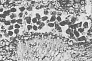
Figs.1 and 2: Angular clots in Ankyropteris
axis cross section, detail from [1], Plate VII, 2 and 5,
interpreted as coprolites from organisms of uncertain identity
[1] and from oribatid mites [2,3], but differently interpreted
here.
Width of Fig.1: 0.5 - 0.57 - 0.63mm. (See
text.) Width of Fig.2: 0.66mm after correction of scales
[1,3].
There are different types of tissue with differential cell
sizes in the Ankyropteris
axis. The clots in Fig.1 are compatible with the cell lumina of the
tissue. Two chains of clots on the left look as if they had not been
dropped there randomly. They look as if they have kept their positions
where they had formed within the cells, a few remaining of which are
seen below left. This suggests that the clots are casts of the cell
lumina left over after the cell walls of the tissue which had been
there where the clots are now had decayed. This idea is supported by
several more observations of this kind. The conspicuously angular clots
in Fig.2 are most probably also casts of cells although no tissue is
seen near them.
Uncertainties of sizes in Figs.1,2 are due to
inconsistencies of size data in [1,2,3]. If 6:1 is true for
the
total view of the Ankyropteris
cross-section in [3], Fig.334, the ratio
for Fig.336 is 24:1, not 12:1, and for Plate VII,5 in [1] it is 35:1,
not 14:1. The captions, too, are erroneous for those two pictures. They
do not show a detail from the main axis but from the mirror image of
the frond stalk in Fig.334.
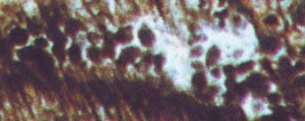
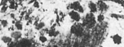
Fig.3:
Angular clots of various sizes in Ankyropteris
cross section, detail
from [3], Fig.335, (which is the mirror image of Plate VII,4 in [1]).
Note the cell above right with a clot of corresponding shape inside. Width of the picture 0.67mm.
The conducting strand of an aereal root of Psaronius seen in
Fig.4 may serve as another example of dark clots representing cell
casts of disintegrated tissue, although the clots are interpreted as
gnawed-away aerenchyma replaced by coprolites in [4].
First, it must be mentioned that the tissue adjacent to the
"star-shaped" xylem strand is no aerenchyma but phloem. Second, the
clot sizes vary strongly, same as the cell sizes of the remaining
phloem. There is even a small patch of well-preserved phloem, seen
above the pocket with the dark clots, with one cell filled with dark
matter, surrounded by empty phloem cells. The compact dark areas in the
phloem are
bounded by straight cell walls nearly everywhere, hence they are no
coprolites but patches of tissue filled with dark matter but not
yet disintegrated.
Note also the separate tiny clot of pentagonal outline.
The phloem between the prongs of the xylem of Psaronius roots is
very
seldom preserved. Hence one may regard the clot formation as a
preservation process for structure data
of tissues prone to rapid decay, namely for shape and size data of the
cell
lumina, which otherwise would get lost.
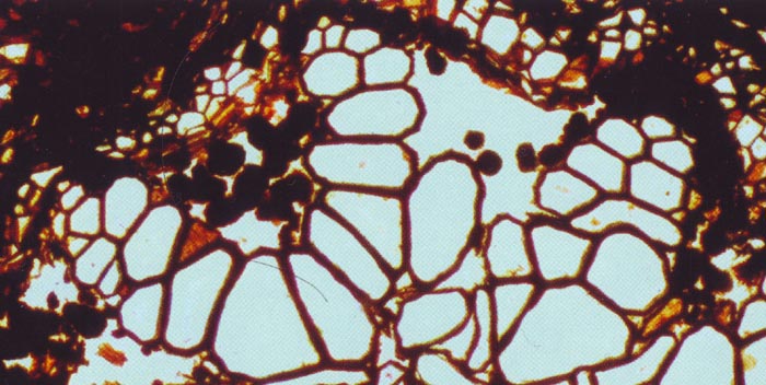
Fig.4: Psaronius
root cross-section, conducting strand with
big tracheids and alleged coprolites replacing aerenchyma according to
[4], here re-interpreted as phloem cell casts. Detail from [4], Fig.8.
Width of the picture about 0.75mm.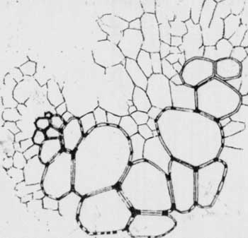
One of the very rare pictures of the phloem in pockets of
the xylem
strand of a Psaronius
root is the drawing in Fig.5 below. An exceptionally well preserved
Psaronius
root conducting strand with clearly seen phloem is presented in [7],
Fig.12, and [8].
Fig.5 (right): Segment of Psaronius
root cross-section, conducting strand with big tracheids
and thin-walled phloem cells. Detail from [5], Plate 40, Fig.13.
Now that ample evidence has been presented supporting the
assertion that probably most of the dark clots in palaeozoic plant
fossils, including coniferous-type wood, and the angular clots in [1-4]
in particular, are no
coprolites but casts of the cell cavities formed in the coherent tissue
and released as the tissue decayed, what remains to be done is to
explain the
nature of the dark matter. A connection to fungus activity has been
proposed in previous treatises, see Misconceptions, Oribatid mite
coprolites. The subsequent disintegration of the tissue after invasion
of the
cells and formation of the dark residue could be due to the ability of
fungi
to break down the cell walls. (Fungi are the only organisms that can
completely break down lignin [6]).
Arguments against the interpretation of angular clots as coprolites in
[1,2,3] had been presented to R.
Rößler since
2007.
The misinterpretations
in [4] have first been noticed by Gert Müller
in 2010. The only response he got was the advice by one of the experts
[4]: "If two professors say the clots are coprolites, you can believe
it."
A critical comment
on [4] (in German) suitable for publication in the journal "Semana" has
been rejected on grounds of pretended copyright violation.
H.-J. Weiss
2011
[1] R. Rößler:
The late palaeozoic tree fern
Psaronius - an ecosystem unto itself.
Rev. Palaeobot. Palyn.
108(2000), 55-74.
[2] R. Rößler:
Between precious inheritance and immediate
experience.
in: U. Dernbach, W.D. Tidwell:
Secrets
of Petrified Plants, D'ORO Publ., 2002, 104-119.
[3] R. Rößler:
Der versteinerte Wald von Chemnitz, 2001, p. 141,169.
[4] M.
Barthel, M. Krings, R. Rößler: Die
schwarzen Psaronien
von Manebach, ihre Epiphyten,
Parasiten
und Pilze. Semana* 25(2010), 41-60.
*( recently re-named, former name: Veröff.
Naturhist. Mus. Schleusingen)
[5] C.G. Stenzel**:
Über die Staarsteine.
**( not to be confused with Sterzel)
Breslau, Bonn 1854,
p.753-893, Plate 40.
[6] T.N.
Taylor, E.L. Taylor, M. Krings : Paleobotany,
Elsevier 2009.
[7] H. Steur,
H. de Kruyk: Psaronius, een boomvaren uit het Laat-Carboon
en het Vroeg-Perm.
Grondboor & Hamer nr.
3/4 2004, 75-82.
[8] H.
Steur:
The tree fern Psaronius,
"Star show". http://www.xs4all.nl/~steurh/
|

|
 6 6 |