Alleged arthropod coprolites re-interpreted
deutsche Version

Several recent attempts to refute the absurd notion of coprolites whose shapes and sizes equal those of the cells eaten by tiny plant eaters has encountered fierce resistance by some of those who interpreted the angular clots in damaged plant tissue as oribatid mite coprolites. (See, for example, "Oribatid mite coprolite sightings – a transient craze ?"  and other contributions accessible from
Chapter "Misconceptions".)
Plain criteria are available for separating the real coprolites from clots erroneously described as such in the scientific literature:

(1) Angular clots are no coprolites.
(2) Clots with shapes and sizes fitting to nearby cells are no coprolites.
(3) Clots inside cells are no coprolites.
(4) Clots arranged in chains and files fitting to the tissue are no coprolites.

By applying these criteria to the published pictures one finds out the misinterpretations rather easily. Some publications with misinterpretations found in this way are listed below, followed by the numbered criteria applying to them:
R.W. BAXENDALE (1979)
K. GOTH, V. WILDE (1992)
C.C. LABANDEIRA, T.L. PHILLIPS, R.A. NORTON (1997)
R. RÖSSLER (2000)
R. RÖSSLER (2001)
R. NOLL, V. WILDE (2002)
D.W. KELLOG, E.L. TAYLOR (2004)
ZHUO FENG, JUN WANG, LU-YUN LIU (2010)
M. BARTHEL, M. KRINGS, R. RÖßLER (2010)
Zhuo Feng, Jun Wang, Lu-Yun Liu, R. Rößler  (2012)
C. Strullu-Derrien, S. McLoughlin, M. Philippe, A. Mørk, D.G. Strullu (2012)
N.A. Jud, G.W. Rothwell, R.A. Stockey (2010)
(1)      
(1) (2) (3)
(1) (2)
(1) (2)
(1) (2) (3) (4)
(3)
(1) (2)
(1) (3) (4)
(1) (2) (3) (4)
(1) (2) (3)
(1) (2) (3) (4)
(1) (2)
[1]
[2]
[3]
[4] 
[5]
[6] 
[7]
[8]
[9] 
[10]
[11]
[12]
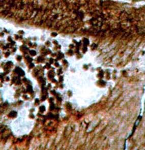
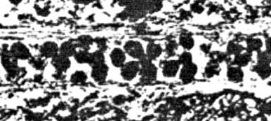
Probably all alleged coprolites are no such in [2, 4, 9-12]. In addition to false coprolites there are clots of uncertain origin in the other papers, and apparently true coprolites in some of them. Among the several pictures of potential coprolites in [1], one shows clots of a different kind:


Cordaites leaf cross-section with angular clotsFig.1: Alleged "small coprolites filling the mesophyll cavity" of a Cordaites leaf from Pennsylvanian coal ball, here interpreted differently. Detail from [1], Fig.9; clot size 30-40µm.


Perhaps the angular shapes of these clots alone would not serve as convincing evidence against their coprolite nature but if compared with quite similar clots in better preserved tissues as seen in pictures in the papers listed above, the following interpretation becomes rather compelling:
The clots had been fills of cells before the cell walls decayed, hence they preserve the shapes and sizes of the cell cavities, or at least their cross-sections. Such idea as an alternative interpretation is virtually absent in the papers on alleged coprolites, hence it is remarkable that it is cautiously mentioned in [1] in connection with the above figure, not in the caption but as a mere half sentence in the subsequent comprehensive discussion:

Quotation: possibly no coprolites       
If this presumption had not been ignored or forgotten, most or all of the oribatid mite coprolite literature would not have been written and would not flourish now, and there would be no need to refute it with critical comments like this one and others in Chapter "Misconceptions".
What had been called, back in 1979, "the degradation of cytoplasm and cell walls" as a possible way of clot formation can be linked to a certain cause now: It is known from the Lower Devonian Rhynie chert that the fungus Glomites rhyniensis invades the small plant Aglaophyton (former Rhynia major), propagates through the intercellular space, eventually intrudes into a cell, branches there profusely to form a dense tangle appearing as a dark clot, thereby feeding on the cytoplasm, sends a hypha through the wall into the next cell to branch there, and so on. (See [10], Figs.3.47 and 3.96.) Clots of such origin may finally fill out the cell so that they represent a replica of its more or less angular interior, as seen more distinctly in another plant in the Rhynie chert, Asteroxylon (Fig.2).angular clots in Asteroxylon
Fig.2: Angular clots
as fills of vaguely visible cortex cells on a cross-section of Asteroxylon: most probably of fungus origin and certainly no coprolites.  Rhynie chert, Lower Devonian, own find.

If inspected carefully, the publications listed above reveal similar evidence contradicting the interpretation of cell-size clots as coprolites in general, and as oribatid mite coprolites in particular.
clots of various sizes and shapes in Ankyropteris, no coprolites
angular clots in Ankyropteris
Figs.3*,4: Alleged coprolites in Ankyropteris, Lower Permian, Chemnitz. Details from [5], Figs.336, 430. Same pictures in [4], Plate VII.


These clots do not look as if randomly dropped into a cavity by some creature. They show a tendency to come in chains as if left over by decaying tissue. Some of them are distinctly angular. Their variation of sizes and shapes is compatible with the structure of nearby tissues. Hence, they are no coprolites.

* Annotation to Fig.3: The captions of the figures in the original publications of which Fig.3 is a detail, Fig.336 in [5] and Plate VII,5 in [4], are erroneous: The figures do not show a detail of the main axis but of the frond stalk. Magnification is not 12:1 but 24:1 in [5], and not 14:1 but 35:1 in [4].

H.-J. Weiss
     Dec. 2010,  2013

[1]  R.W. BAXENDALE: Plant-bearing coprolites from North American Pennsylvanian coal balls.
      Palaeontology 22(1979), 537-548, plates 65,66.
[2]  K. GOTH, V. WILDE:  Fraßspuren in permischen Hölzern aus der Wetterau.
      Senckenbergiana letaea 72(1992), 1-6.
[3]  C.C. LABANDEIRA, T.L. PHILLIPS, R.A. NORTON: Oribatid mites and the decomposition
      of plant tissues in paleozoic coal swamp forests.  Palaios 12(1997), 319-53.
[4]  R. RÖSSLER: The late palaeozoic tree fern Psaronius  -  an ecosystem unto itself.
      Rev. Palaeobot. Palyn. 108(2000), 55-74.
[5]  R. RÖßLER: Der versteinerte Wald von Chemnitz, 2001, p 141,155,169.
[6]  R. NOLL, V. WILDE :  Conifers from the „Uplands“ – Petrified wood from Central Germany. 
      in: U. DERNBACH, W.D. TIDWELL : Secrets of Petrified Plants, D'ORO Publ., 2002.
[7]  D.W. KELLOG, E.L. TAYLOR: Evidence of oribatid mite detrivory in Antarctica during the
      Late Paleozoic and Mesozoic.  J. of Paleontology 78(2004), 1146-53.
[8]  ZHUO FENG, JUN WANG, LU-YUN LIU: First report of oribatid mite (arthropod) borings and coprolites
      in Permian woods from the Helan Mountains of northern China.
      Palaeogeography, Palaeoclimatology, Palaeoecology 288(2010), 54-61.
[9]  M. BARTHEL, M. KRINGS, R. RÖßLER: Die schwarzen Psaronien von Manebach, ihre Epiphyten,
      Parasiten und Pilze.  Semana 25(2010), 41-60.
[10] T.N. TAYLOR, E.L. TAYLOR, M. KRINGS:  Paleobotany, Elsevier 2009.
[11] Zhuo Feng, Jun Wang, Lu-Yun Liu, R. Rößler: A novel coniferous tree trunk with septate pith ...
     Int. J. Plant Sci. 173(2012), 835–848.

[12]  C. Strullu-Derrien, S. McLoughlin, M. Philippe, A. Mørk, D.G. Strullu: Arthropod interactions with bennettitalean roots ...
      Palaeogeography, Palaeoclimatology, Palaeoecology 348–349(2012), 45-58.
[13]   N.A. Jud, G.W. Rothwell, R.A. Stockey: Paleoecological and phylogenetic implications of Saxicaulis meckertii ... :  
      Int J. Plant Sci. 171(2010), 915-25.
Annotation: A critical comment on [9] (in German) suitable for publication in the journal "Semana" has been rejected on grounds of pretended copyright violation.
quartz crystal with wood inside
Fossil Wood News 7

Site map
Fossil Wood News
Wood rot or coprolites
Silicified wood