Alleged coprolites of "unknown creatures" replace
alleged oribatid mite coprolites
After the row about oribatid mite coprolites in Palaeozoic plant
fossils had calmed in the latter years, the attention paid to the
subject has unexpectedly increased again. It started in 2010 with a
paper by
Zhuo Feng et
al. [1], recognized and described as erroneous in [2].
(See also Misconceptions.)
As a novelty, in a publication by M. Barthel,
M.
Krings, R. Rößler [3]
the dark granules are no more assigned to the favourite oribatid mites
as done before in several papers by various authors (see compilation in
[4])
but to "unknown creatures". The reader is also told that fungi are
closer related to animals than to plants. Is this intended to pave the
way for a recognition of the little clots as fungus formations ? This
would be a too optimistic interpretation. It will rather take some
effort to remove the absurd notion of coprolites of small plant eaters
whose shapes and sizes, including edges and angles, agree with those of
the cells eaten. No
effort is needed to convince average people but this is different with
the palaeobotanists who promoted the mite coprolite hypothesis.
Therefore, the old arguments have to be applied here to the new
pictures.

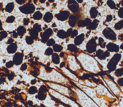
Fig.1: Part of Fig.16 in [3], there interpreted as "coprolites in the
conducting tissue (scalariform
tracheids) of a Psaronius
root", here interpreted differently.
(*)
According to experience, small clots of this kind agree with the cells
of the "eaten" plant tissue with respect to shape, size, and
variability.
Contrary to the assertion in [3], no clot is seen inside the tracheids.
All of them are lying around outside. They are angular like the cells
of the decayed tissue whose remains are seen in the corner below left.
As in numerous similar cases, they can easily be interpreted as fills
of cells whose walls have gone. In this connection, the next picture is
revealing.

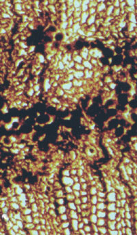
Fig.2 (right): Detail from Fig.17 in [3],
there interpreted as "Dadoxylon
sp cross-section of secondary xylem with coprolites within a gallery", here
interpreted differently. (*)
Where is a gallery in this picture ? No such is seen but there are
other things worth being noticed: individual dark clots inside cells,
chains of dark clots, and narrow dark pith rays. There is a pith ray
with dark fill, kinked into a conspicuous Z-shape.
These observations are compatible with the notion of something
propagating from cell to cell, or also to more distant cells, to
produce dark fills. Such phenomenon is known from the fungus Glomites
rhyniensis: [6]
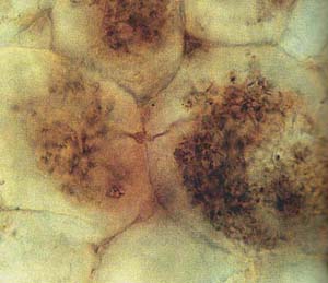
Fig.3.47. It enters into the cell, branches profusely
to form a tangle of very thin hyphae,
filling the cell more or less completely and looking like a dark clot,
then penetrating the wall to get into the neighbouring cell
to branch there, and so on. (See picture below.) 
Fig.3: Picture from [5] Fig.19 and [6] Fig.3.96: Dark
clots consisting of a tangle of very thin hyphae of Glomites in cells
of Aglaophyton
from the Lower Devonian Rhynie chert.
Note the hypha penetrating the wall.
To sum up, one can state that the alleged coprolites are neither
related to oribatid mites nor to unknown creatures but are no such at
all, same as in numerous other cases of small dark clots in silicified
plants.
* The misinterpretation in [3] was noticed by Gert Müller
(Dresden).
For more critical annotations to [3] see "Wood rot"
and "Remnants".
H.-J.
Weiss
Dec. 2010
[1]
Zhuo Feng, Jun Wang, Lu-Yun Liu :
First report of
oribatid mite (arthropod) borings and coprolites in Permian woods from
the Helan Mountains of northern China.
Palaeogeography,
Palaeoclimatology, Palaeoecology 288(2010), 54-61.
[2]
H.-J.
Weiss:
Dubious oribatid mite coprolites once more: Comment on Z.
Feng et al. (2010),
[3]
M. Barthel, M. Krings, R. Rößler: Die schwarzen Psaronien
von
Manebach, ihre Epiphyten, Parasiten und Pilze. Semana 25(2010), 41-60.
[4]
H.-J.
Weiss:
Oribatid mite coprolite sightings – a transient craze ?
[5] H.
Kerp: De Onder-Devonische Rhynie Chert ... . Grondboor
& Hamer 58(2004), 33-50.
[6] T.N.
Taylor,
E.L. Taylor, M. Krings : Paleobotany, Elsevier 2009.
Annotation (2011): The authors [3] were not interested in discussing the problem.
A critical comment
on [3] (in German) suitable for publication in the journal "Semana" has
been rejected on grounds of pretended copyright violation.
Annotation
(Sept. 2016): Years of continuing own effort to unsettle the proponents
of the coprolite interpretation of clots in petrified wood have not
been in vain. Finally they accepted the fungus interpretation proposed
above: Another investigation [7] revealed the presence of several
different fungi in the very same Psaronius specimen as in [3]. Even hyphae penetrating the cell wall as shown in Fig.3 have been found with this Psaronius
specimen. Unfortunately, this laudable change of mind of two of the
authors [3,7] comes along with the reluctance to admit errors. The
conspicuous cell-size clots erroneously offered as coprolites in [3]
are not mentioned at all in [7]. Hence this error, like several others
in palaeobotany, has to be addressed expressly.
[7] M. Krings, T.N. Taylor, C.J. Harper, M. Barthel: Endophytic fungi in a Psaronius root mantle from the Rotliegend of Thuringia, Germany.
87th Annual Conference Paläontologische Gesellschaft, Dresden, Sept. 11-15, 2016.
Annotation
(2017): A detailed paper [8] on the same fossil confirms the previous
annotation. Misleadingly, it claims to "complement" [3] without
mentioning that [3] favours the erroneous interpretation of fungus clots as coprolites. That error had been rejected since 2007, also by the above contribution in 2010, and several more times since.
[8] M. Krings, C.J. Harper, J.F. White, M. Barthel, J. Heinrichs, E.L. Taylor, T.N. Taylor:
Fungi in a Psaronius root mantle from the
Rotliegend (Asselian, Lower Permian/Cisuralian) of Thuringia,
Rev. Pal. Pal. 239 (2017), 14-30.
|

|
 4 4 |

 4
4


 4
4