Varied nematophytes in Rhynie
chert
Recent finds of nematophytes in the Rhynie chert,
notably hitherto unknown
ones, provide information which may help to demystify these "Enigmatic
Organisms" [1]. Nematoplexus
[2] is the only one known to consist of screw-like
wound tubes. The comparison with a
screw is appropriate since the tubes are always wound in a right-handed
way, with a definite pitch of the thread. Contrary
to fossil evidence, the "spiral coils" have repeatedly been ascribed
to all nematophytes [3,4], which is clearly erroneous.
The tubes of Nematoplexus
from
own finds are larger than the original ones from [2]: Rhynie
Chert News 29.
Unexpectedly,
straight or weakly curved tubes, too, have
been found associated with
Nematoplexus
(Fig.1). As a remarkable fact, there
are no tubes with shapes intermediate
between the straight or weakly curved ones and
the screw-like ones: Rhynie
Chert News 51. Among the confusing tangle of tubes, several
turns of one screw as in
Fig.1 (right) are not often seen. 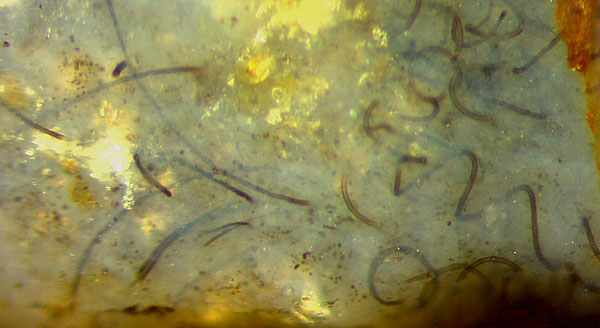
Fig.1: Nematoplexus
with slightly curved and screw-like wound tubes, width 11-12µm.
Image width 1.5mm.
The term
"branch-knot" and its repeated use in connection with tube generation
has led to the view that it is a dense tangle of tubes where the
tubes profusely branch inside before they venture out. This notion has
been refuted in Rhynie
Chert
News 134,
136,
152.
To
avoid consolidation of erroneous views, the branch-knots may simply be
called clots. They are no tangles of tubes but bodies of their own with
a (not clearly defined) surface. The tubes arise from that surface or near-surface region.
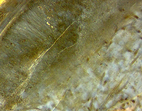
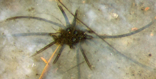 No clot related to the
slightly curved tubes as in Fig.1 (left) has been found yet
but, as a big surprise, a bunch of slightly
curved big tubes attached
to a big clot (Fig.2) provides another enigma: Slightly
curved
big tubes have only been found near the "normal" screw-like
ones as if they were
mutually related somehow:
See Rhynie
Chert
News 135,
137.
It
appears that even the seemingly thoroughly inspected Nematoplexus
still hides big secrets.
No clot related to the
slightly curved tubes as in Fig.1 (left) has been found yet
but, as a big surprise, a bunch of slightly
curved big tubes attached
to a big clot (Fig.2) provides another enigma: Slightly
curved
big tubes have only been found near the "normal" screw-like
ones as if they were
mutually related somehow:
See Rhynie
Chert
News 135,
137.
It
appears that even the seemingly thoroughly inspected Nematoplexus
still hides big secrets.
Fig.2: Slightly curved
big tubes, 17-22µm, and a few very thin ones, 4-5µm, emerging from a
big clot;
nearly straight thin tube, 9µm, passing by. Same
scale as Fig.1, image width 1.3mm.
Since Nematoplexus
is apparently the only nematophyte
with screw-like wound
tubes, the aspects of all others lately found in
the Rhynie chert differ
greatly. An impressive example of a stack of nearly straight aligned
tubes with large diameters is seen in Fig.3.
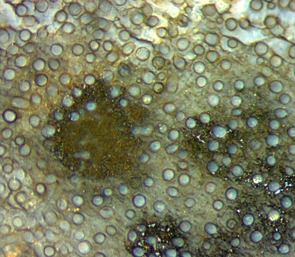
Fig.3: Aligned big tubes of unknown nematophyte,
up to 70µm across.
Same scale as Figs.1,2, image width 1.5mm.
A
few conclusions can be drawn from details in Fig.3: The absence of
mineral debris between the tubes seems to indicate that they did not
grow separately in the swamp water which became the Rhynie
chert
but rather in a lump of organic gel produced by themselves to live
in. This
would be compatible with the irregularities
seen above, which are possibly due to beginning decay, and with the
beginning
formation of a pseudo-cell pattern of shrinkage cracks in the gel below
left: Rhynie
Chert
News 30.
Also the different aspect of the chalcedony inside and outside the
tubes indicates different conditions of silicification, as discussed in
Rhynie
Chert
News 154.
The
tiny precipitates seen
as deposits inside two
tubes below right must have formed
while the tube content was fluid. Hence, it
had decayed before silicification but the organic gel surrounding the tubes had not, keeping the tubes apart until
silica gel stabilized the structure. Note that there are no tubes touching.
Another nematophyte shows a rather different structure: Fig.4. The chaotic arrangement of limp tubes seems to indicate a liquefaction of the organic gel before silicification but this interpretation is questionable.
Inside
the lump of gel, the tubes are protected against exsiccation and
microbial attack: Rhynie
Chert
News 155.
Most
probably, other nematophytes,
too, made use of this two-fold protection.
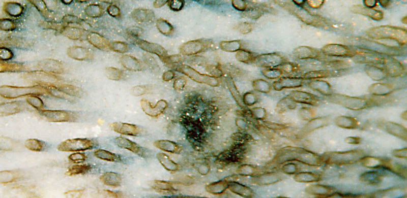
Fig.4 (left): Unknown nematophyte
with big tubes up
to 60µm across, poorly aligned, irregularly curved. Same scale as
above, width 2mm.
Gel turned into bluish
chalcedony has drawn the attention to
surprisingly narrow nematophyte
tubes (Fig.5), which hitherto may have been overlooked or
misinterpreted as less interesting fungus hyphae: Rhynie
Chert
News 86.
Yellow mineral debris is seen above left and through the translucent
gel on the right.
Fig.5 (below): Unknown nematophyte
with very narrow tubes about
6-8µm across, irregularly curved. Same scale as above, width 1.4mm.

A clearly defined outline is missing with the nematophytes in
Figs.1-5, where the lumps of gel with tubes inside are seen as
fragments in the chert. Nematophytes
with preserved outline
are shown in other samples: Rhynie
Chert News 99.
The pale spots of densely spaced tubes in
Fig.6, below left and above right, might be the expected centres of
tube formation. With
tube diameters ranging from well below 10µm up to 27µm and a width of
the whole organism of hardly more than 2cm, this nematophyte resembles
the flat one in Rhynie Chert
News 46 but it
lacks the clearly structured outer sheath seen on that specimen.
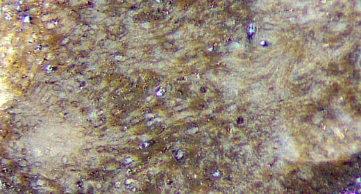
Fig.6 (left): Unknown nematophyte with tubes up
to 27µm across. Same scale as above, width 1.3mm.
Fig7. (below): Nematophyte with layered structure and different
types of tubes in the layers,
thus resembling Nematophyton
taiti. Same
scale as above, width 1.5mm.
The nematophyte
in Fig.7 is the only one among the own finds with distinctly different
types of "tissue", as outlined in
Fig.8. See also Rhynie Chert
News 35, 46, 153.
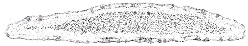
Fig.8: Attempted reconstruction of the nematophyte in Fig.7. Width
3.5cm.
Lately, the similar Nematophyton
taiti
has been assorted among the ascomycetes and
renamed Prototaxites
taiti [5]. Possibly the present fossil and other
nematophytes, too, will have to be reconsidered.
The detail in Fig.7 has been chosen such that it is reminiscent of the
outer sheath of Pachytheca.
That
spherical nematophyte
is known from several locations but one
exceptionally preserved specimen
has been found in the Rhynie chert:
Rhynie
Chert News 1, 36, 44,
(No picture included here.)
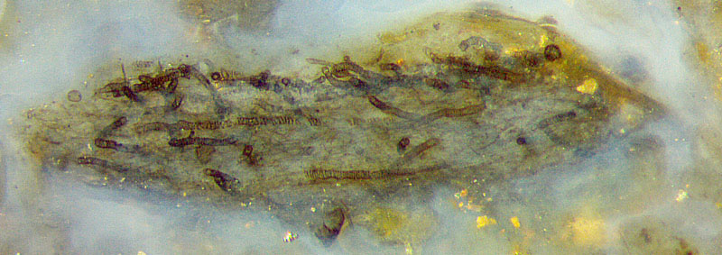
Tubes with patterned walls occasionally seen among "normal" ones with Nematoplexus seem to be "out of place" there but are normal with Nematothallus:
Fig.9. Their obvious similarity with tracheids has given
rise to reasonable suggestions concerning the evolution of land plants [6] but also to quite absurd ideas about water flow in cells [7].
Fig.9:
Worm-like tubes about 20µm wide with patterned walls, tiny tubes (<3µm ?) in
between, apparently grown in a lump of organic gel, possibly first Nematothallus in 3D-preservation.
Width 2mm, same scale as Figs.1-7.
To sum up, the nematophytes lately found in the Rhynie chert and
briefly presented in Figs.1-9 provide evidence that ...
- the only
nematophyte with
screw-like wound tubes, the allegedly well investigated Nematoplexus,
remains
highly enigmatic with its apparent affiliation to hitherto unknown structures
(Figs.1,2),
- the clots called "branch-knots" are no tangles of
branching tubes but lumps producing the
tubes in obscure ways (Fig.2),
- there are several hitherto unknown nematophyte forms and species
in the Rhynie chert (Figs.1-8),
among
them species with remarkably narrow or wide tubes.
- Nematoplexus, is not, as once assumed and numerous times repeated [1], a permineralised form of Nematothallus.
Samples shown in Figs.1-9:
Rh9/86.1, Rh15/79.1, Rh2/81.1, Rh13/7.1, Rh3/9.1, Rh13/1.2,
Rh2/7.6.
H.-J.
Weiss 2020
[1]
T.N. Taylor,
E.L.Taylor, M. Krings: Paleobotany, Elsevier 2009.
[2] A.G.
Lyon: On the fragmentary remains of an organism
referable to the Nematophytales,
Trans. Roy. Soc. Edinburgh
65(4)(1962): 79-87.
[3]
P. Selden, J. Nudds:
Evolution of fossil ecosystems. Manson publ. 2012, p.84.
[4] www.abdn.ac.uk/rhynie/nemato.htm
[5]
R.
Honegger, D. Edwards, L. Axe, Ch. Strullu-Derrien:
Fertile Prototaxites
taiti: a basal ascomycete with
inoperculate, polysporous asci lacking croziers.
Phil.Trans. Roy. Soc. B 373 (2017): 20170146.
[6] P.K. Strother: Clarification of the genus Nematothallus Lang: J. Paleont. 67(1993), 1090-1094.
[7] K.J. Niklas, V. Smokovitis: Evidence for a conducting strand in early Silurian plants, (1983).
 |
 |
156 |