More nematophytes in the Rhynie chert
Two
species of the enigmatic "filamentous plants" had been discovered in
the Rhynie chert and described as Nematophyton
(1921) and Nematoplexus
(1962).
Another nematophyte, Pachytheca,
known for a long time from other
locations, had been found at Rhynie in 2003 and
described in Rhynie
Chert News 1.
More nematophytes have been found since: a flat
one (2010) resembling but differing from Nematophyton, one
of uncertain overall shape but with much bigger tubes (2005), another one
with spirally coiled
tubes (2009) but differing
from
Lyon's Nematoplexus
in
more than one way, a similar one showing monstrous "branch knots" of
slightly curved big tubes (2015). 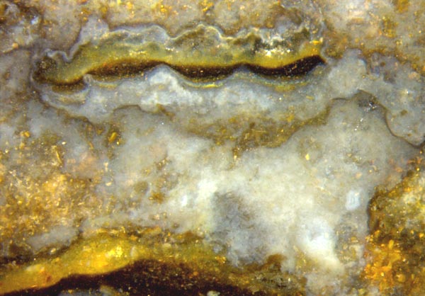
A
quite different aspect is offered by randomly arranged very thin and
inconspicuous tubes or fragments thereof. Their variable curvature
distinguishes them from aquatic fungus hyphae whose straight
sections make a different aspect in the chert.
The tubes are occasionally
seen scattered within pale or white clouds in the usually darker
chalzedony, suggesting the idea of a nematophyte connection. The
idea was not pursued for the scarcity of fossil evidence until the
tubes appeared abundantly on the cut faces of an olive-brown chert
sample (Fig.1).
Fig.1 (right): Rhynie chert with cloud-like
formations, apparently former silica gel slightly older than other
precipitates
and deposits. Note the remains of small tubes faintly seen in a lump
below the
middle. Width of the picture 8mm.
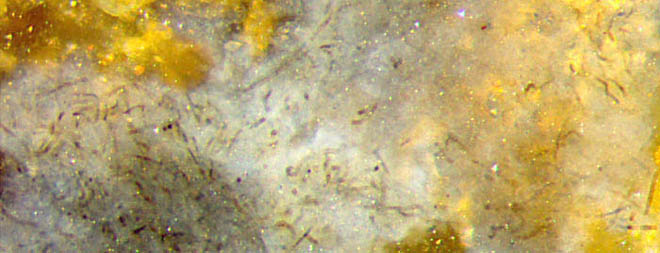
Fig.2
(above left): Randomly distributed tube fragments restricted to a part
of
chert appearing as a cloud. The cloud is thin on the right
so that the yellow chert behind is shining through.
Width of
Figs.2-6:
1.4mm.
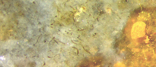
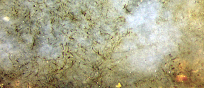
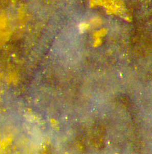
Fig.3 (above left): Similar as Fig.2, mineral
debris on the
right.
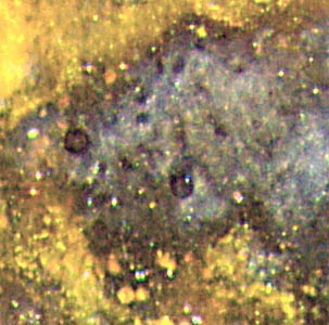
Fig.4 (above right):
Similar as
Fig.2, no tubes in part of the cloud.
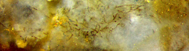
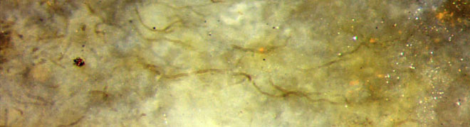
Fig.5 (left): Similar as Fig.4.
Fig.6 (right):
Sample surface, longer tubes visible.
From
the observation that the cloud-like areas seen on the surface and cut
faces of some chert samples are free from the mineral precipitates
abundantly present around them (Figs.1-3) it can be concluded that the
clouds formed as lumps of gel before mineral silt formed in the water.
The observation that tubes like those in the above pictures are
restricted to the interior of the said clouds hints at a deeper
connection. In the absence of additional information, several options
are thinkable. The clouds could have been microbial colonies held
together by organic slime or gel, then invaded by a tube-like organism
feeding on them. As another possibility, the tubes could have produced
the gel in the same way as nematophytes are supposed to do. Organic
gel could have triggered the formation of silica gel
and become replaced by the latter.
Alternatively,
substances released by microbes or tubes could immediately have caused
silica gel formation by changing the acidity of the water.
Most often
the light-coloured clouds in chert seem to be "clean". This could mean
that they had never been inhabited by tube-like organisms, or the
latter are
decayed and no more seen. This possibility is suggested by the
observation of very faint remains of thin tubes in some chert samples.
There
does not seem to be an easy interpretation of this type of fossil. The
overall aspect of the tangle of randomly distributed irregularly curved
tubes resembles that of unnamed nematophytes described in Rhynie
Chert News 13,
35
except
for the very small tube diameters of 6-8-(10)µm in this case, compared
to
50-70µm and 25-30µm of the said nematophytes. Also the tubes in the
above pictures seem to be less densely spaced. This, however, might be
due to fast decay of part of the tubes, which could also explain the
observation that there were lots of tube fragments. Tubes are seldom
seen extending over some distance as in Fig.6.
Although a
similarity to nematophyte tubes is obvious, the apparent absence of
so-called "branch knots", a typical but not yet understood feature of
nematophytes, raises doubts concerning a nematophyte affiliation. As an
additionally confusing fact, this sample contains also a few tubes of
different types whose possible relation
to the tubes in the above pictures is questionable but cannot be ruled
out (Figs.7,8).
Figs.7,8: Some of the very few big tubes in this sample,
among narrow ones of about 7µm as in Figs.2-6,
diameters about 30µm (with rings on the wall) and 20µm, in bluish
chalzedony surrounded by mineral debris.
(The white dots are not
relevant.)
Width of the pictures 0.3mm,
magnification twice as large as in Figs. 2-6.
The
very few big tubes with diameters of about 15, 20, 30µm are seen
together with some narrow ones in
smaller patches of bluish chalcedony separate from the larger whitish
clouds. The combination of different types of tubes, including those
with rings on the wall, is known from nematophytes. If the tubes in
Figs.7,8 are nematophyte tubes, which they probably are, one may
conclude that the narrow tubes in Figs.2-6 are nematophyte tubes, too.
Hence, what has been described here is probably a new species of the
enigmatic nematophytes which is not rare in the Rhynie chert but is
easily overlooked.
The tubes have been discovered in 2015 on this sample Rh3/9 found in 1998.
H.-J. Weiss
2015
 |
 |
86 |