Tubes at Nematoplexus
"branch-knots"
 Since
its discovery [1], new finds of this "enigmatic organism" [2] known
from the Rhynie chert only have
provided new problems in addition to a few answers to old
questions. Apparently it is either unbelievably
versatile as a species or it is nearly always associated with one
or more other
nematophyte species. Anyway, Nematoplexus
is very peculiar, as indicated by this picture.
Since
its discovery [1], new finds of this "enigmatic organism" [2] known
from the Rhynie chert only have
provided new problems in addition to a few answers to old
questions. Apparently it is either unbelievably
versatile as a species or it is nearly always associated with one
or more other
nematophyte species. Anyway, Nematoplexus
is very peculiar, as indicated by this picture.
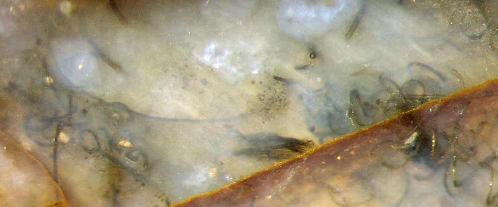
Fig.1: Nematoplexus
tubes:
spiralling ones (10-12µm) near two obscure "knots" on the right, a
bunch of straight ones below the middle, a long bow dimly
seen in the
depth, and broader slightly curved ones (18µm) above.
Image width 2mm.
The various aspects of Nematoplexus tubes
evident from own chert finds have been described in Rhynie
Chert News 29,
51,
102,
134, 137.
The present contribution is meant to consider the
so-called
branch-knots in a wider context. Usually they are surrounded by a
tangle of tubes so the
very "knot" is not seen. Since the conspicuous
tubes are never seen branching, the
simple idea had come up that there might be profuse branching
inside
the knots. This seemed to be compatible with the questionable statement
in [1], p.80, that "tubes can be seen entering and leaving them." Hence
the
term "branch-knot" was
accepted as if it could explain how the tubes are
generated.
It
is probably not relevant but must be mentioned here that the "knot" in
Fig.1 is peculiar as it is really two knots near each other. The
upper one is traversed by a big straight crack, which has provided
the rare opportunity to
illuminate half the knot
with reflected light from behind such that a rare sight has become
visible in
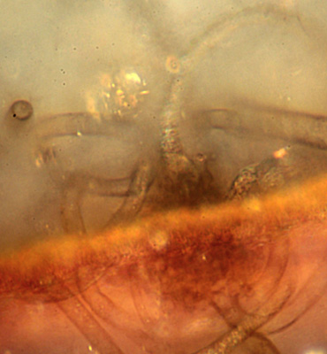
Fig.2: A kind of
foot of a spiralling tube below right is seen connected to a kind of
rugged surface of the central
body of the "branch-knot". Another
spiralling tube apparently
begins on the dark side
of the divide with a
broad foot, too.
Fig.2: Nematoplexus
"knot", detail of Fig.1 with appropriate
magnification,
illumination, and focus
depth.
Image
width 0.2mm. Photograph by G. Schmahl.
Even though Fig.2 may seem rather confusing, it
provides valuable information: The "branch
knot" is not simply a dense tangle of spiralling tubes but a
separate clot with a rugged surface of granular appearance, where the
spiralling tubes start from, without any branching. If there is
some kind of branching unseen inside, it does not produce the
emerging tubes.
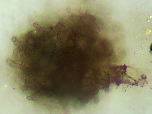
Fig.3: Nematoplexus
"knot" with 4µm-tubes and
only one stump of the spiralling 12µm-tubes left.
Image width 0.11mm. Photograph
by G. Schmahl.
The
globular shape of the central clot may become clearly visible when the
spiralling tubes decay and
vanish, as with the clot in Fig.3, seen amidst
decaying 12µm-tubes in
Rhynie
Chert News 133.
This clot seems to be similar to the one in Fig.2: no
smooth surface, here with short (?) 4µm-tubes poking out. One
4µm-tube is also seen in Fig.2. Probably
the 4µm-tubes have mostly
been overlooked but on
some clots they are absent: Rhynie
Chert News 136.
The considerable diversity of Nematoplexus as
indicated by Fig.1 is even topped by the big variant described
in Rhynie
Chert News 135. Big tubes with a club-like broadened "foot" seem to stick to a
clot with a fuzzy surface
(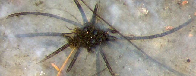 Fig.4). The
big tubes are of the same type as the 18µm-tubes in
Fig.1. The picture plane apparently cuts through the clot near its
surface, revealing circular sections of some tube "feet", up to 34µm: Rhynie
Chert News 135,
there Fig.3. Also seen is an irregular structure inside the clot but no branching tubes.
Fig.4). The
big tubes are of the same type as the 18µm-tubes in
Fig.1. The picture plane apparently cuts through the clot near its
surface, revealing circular sections of some tube "feet", up to 34µm: Rhynie
Chert News 135,
there Fig.3. Also seen is an irregular structure inside the clot but no branching tubes.
Fig.4: Uncommon
Nematoplexus
"knot" with slightly
curved tubes
of 17-22µm and 4-5µm,
one
separate 9µm-tube of unknown affiliation; crack stained with
hematite.
Image
width 1.3mm, same scale as Fig.1.
For more uncommon Nematoplexus clots see Rhynie
Chert News 126,
Finally it
appears that the statement in [3] that "Branching
of the tubes occurs in
...very tightly coiled knots of tubes showing repeated and closely
spaced branching" does not apply to the conspicuous
tubes seen here, spiralling or not. They
do not branch, neither inside nor elsewhere. Some
of the clots are so small that there would not be enough room for "tightly
coiled repeatedly
branching" tubes: Rhynie
Chert News 136,
there
Fig.3.
The idea of "tubes ...
entering and leaving" the knots [1] is misleading. Apparently the tubes
come from the surface or
near the surface of the
clot, where they possibly are in contact with an internal structure possibly
involving very thin tubes
of a different type [1]. However, no
tubular structures have been seen inside the
above clots.
The
question concerning the origin of the tubes has been partially answered
here but other questions have remained unanswered: Why do the tubes of Nematoplexus vary
tremendously even in one chert sample:
straight, curved, and spiralling ones with several diameters,
smooth-walled and patterned ones ?
Sample: Rh14/35: Fig.3;
Rh15/79: Figs.1,2,4.
H.-J.
Weiss 2020
[1] A.G. Lyon:
On the fragmentary remains of an organism referable to the
nematophytales, from the Rhynie chert, Nematoplexus rhyniensis.
Trans. Roy. Soc. Edinburgh
65(1961-62), 79-87, 2 tables.
[2] T.N. Taylor,
E.L.Taylor, M. Krings: Paleobotany, Elsevier 2009.
[3] www.abdn.ac.uk/rhynie/nemato.htm
 |
 |
152 |