Nematothallus
and Nematoplexus
in one chert sample
This fossil resembles
the nematophyte Nematothallus,
which had been
found as compresssions only but never
in chert. The
similarity has
been pointed out by P.K. Strother
[1,2], hence it is called Nematothallus
here, for
simplicity.
It is also shown in Rhynie
Chert News 107
and 122.
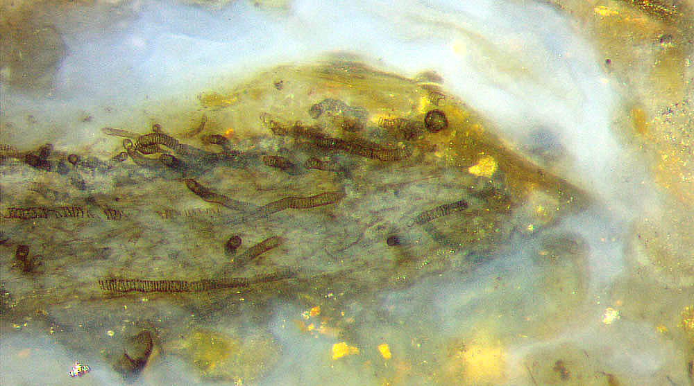
Fig.1:
Formerly separate fragment of gel with Nematothallus,
here
in bluish chalcedony from silicified water of the swamp containing Nematoplexus
(see Fig.4).
Image width 1.38mm.
The unforeseen
discovery of Nematothallus in
a
chert sample incidentally containing the rare fossil
Nematoplexus
seemed to support the
assumption that Nematoplexus
"may represent the permineralized equivalent of Nematothallus" [3].
This is
one of the reasons why the disputed nematophytes
are considered here once more.
The statement in [4] that "Nematophytes
appear to generally comprise networks of intertwined spirally coiled
tubular cells" is not true.
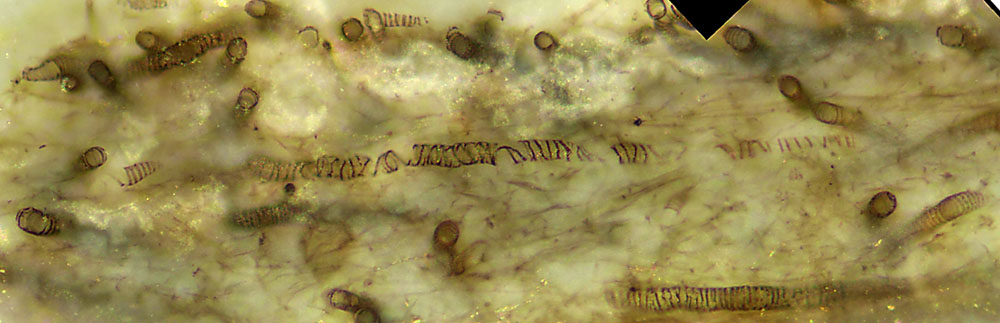
Fig.2 (left): Nematothallus, detail
from Fig.1: tube wall patterns consisting of rings and spiral
fragments of 19-23µm diameter. Image
width 0.97mm.
Photograph by G. Schmahl.
The lump in Fig.1
seems to have separated from a bigger lump or layer of Nematothallus
in gel and got near Nematoplexus
also grown in that habitat. All together silicified
such that the two nematophytes are found now within a mutual distance
of mere 3cm.
Part of the left end of the Nematothallus lump
is seen magnified in Fig.3.
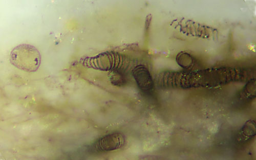
Fig.3: Nematothallus parts
of various aspect, enlarged part of Fig.2 above
left.
Image width 0.3mm. Photograph
by G. Schmahl.
A few details are worth mentioning: Not
the tube walls but the reinforcing patterns have been preserved. The
former
diameter of the tubes had probably been about the same size. Conspicuous
is the variable diameter of one of the tubes.
As seen above right, the tube wall pattern is not always ring-like or
spiral-like but can be rather rugged. The
nature of the bubble
on the left is not known.
A narrow tube, diameter
about 4µm, indicates that there may be more hidden structure. Tubes
of 1.5-3µm among
the big ones have been reported from Nematothallus
compressions [1,2], and
possibly the thin line fragments in Figs.1,2 are
the remains thereof. Small
tubes with sizes of 3µm and below, in addition to the larger ones, are
also found close to Nematoplexus "branch
knots", as seen in Rhynie
Chert News 133
.
Despite of these similarities it
is not justified to regard Nematoplexus
as a form of Nematothallus.
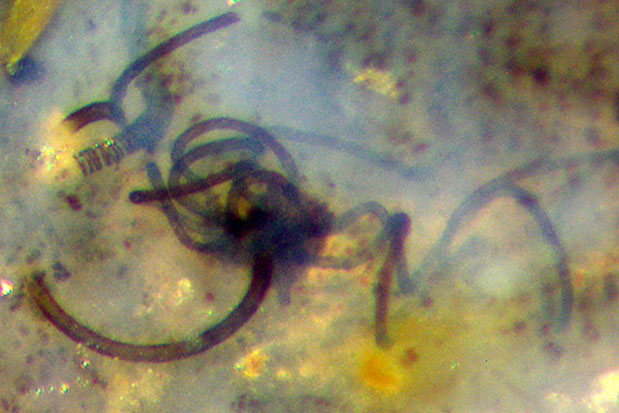
Fig.4 (right): Nematoplexus
"branch knot" with two types of spiralling tubes emerging, diameters
10µm and 16µm, also one patterned tube of dubious affiliation, 24µm.
Image width 0.6mm, same scale as Fig.2.
The conventional term "branch knot" in the
literature on Nematoplexus
suggests an
origin of the tubes by branching. The
contribution Rhynie
Chert News 134
(there Fig.2) favours a different view: There, two
10µm-tubes, each one with a slightly broadened "foot", seem to
stick to the poorly defined surface of a central clot with about 75µm
diameter. There is no indication that the tubes had been produced by
branching inside the clot. In this Fig.4, the central clot of the
"knot" is
of about the same size. Although its surface is less well defined, it
can be assumed that here, too, the tubes emerge from the surface. Also
there would hardly be enough room for many branching tubes
inside.
Near the knot in Fig.4,
the tubes have not yet formed their ideal
spiral shape.
The
bigger tube in Fig.4 emerges from the depth below the
knot in a half circle towards
the
cut plane where it is cut off and hence no more seen, then dives down
below the cut plane above left and thus is in the picture again. Nearby
there is
a big tube with patterned wall,
which
looks surprisingly similar to the Nematothallus tubes
in the above pictures.
(For easy comparison,
Figs.2 and 4 have got the same scale.) It
might
be a stray relic from decayed Nematothallus,
or else it could be one of the less abundant patterned tubes seen
emerging from "branch
knots" of Nematoplexus
[4,5].
As
a peculiar fact, the patterned tubes in these pictures are transparent,
without a visible wall, but the
spiralling tubes from
the
same sample (Fig.4)
appear dark, with more or less translucent
well-defined walls.
Even though the
suggestion in [3] that Nematoplexus
"may represent the permineralized equivalent of Nematothallus"
is not at all substantiated, intriguing questions remain:
Is there a reason why it is not unlikely that the two rare nematophytes
are together in one piece of chert ?
Why are the worm-like tubes with patterned wall
occasionally emerging from
Nematoplexus "branch
knots" surprisingly similar to those of Nematothallus ?
Sample: Rh9/86, 0.28kg, 2003.
H.-J.
Weiss 2020
[1]
P.K. Strother: Clarification of the genus Nematothallus Lang: J.
Paleont. 67(1993), 1090-1094.
[2] W.H. Lang: On the
plant-remains from the Downtonian ... . Phil. Trans. Roy. Soc. London B
227(1937), 245-291.
[3] T.N.
Taylor,
E.L.Taylor, M. Krings: Paleobotany, Elsevier
2009. (Scale
error on Fig.6.9: scale bar not 100µm, rather 20µm ?)
[4] www.abdn.ac.uk/rhynie/nemato.htm
[5] Rhynie
Chert News 137
 |
 |
151 |