Nematophyte big tubes with
precipitate
Nematophytes with tubes wider than those of Prototaxites
are worth being noticed even without the rare phenomenon of mineral
precipitates seen in some of the tubes as in Figs.1,2. The
tube widths of Prototaxites are said to be
below 50µm but are usually pictured as about 25µm [1,2]
while those of this specimen reach 65µm.
The yellow and red precipitates most probably consist of iron oxides.
Since the tiny grains are seen here settled at the bottom of
the
tube sections, it can be concluded that they had formed while the
interior of the tubes was fluid. The transient
existence of a fluid
fill of the tubes is unexpected since they
had been filled with protoplasma while alive, and with silica gel while
becoming silicified.
Apparently,
the decaying cell content became watery and its silicification was
delayed inside because the still present cuticle on the tubes hindered
the diffusive inflow of silica.
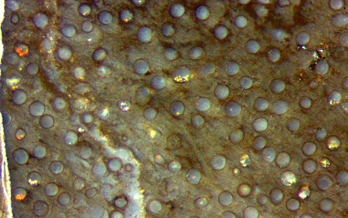
Fig.1: Cross-section
of unknown nematophyte in Rhynie chert, with well aligned tubes, width
up to 65µm, with mineral precipitates at the bottom of a few tube
sections. Width of the image 1.4mm.
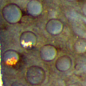
Fig.2: Detail of Fig.1 below left.
Width of the image 0.3mm.
There is no indication why the precipitates
did not form in every
tube or outside. Perhaps it is another manifestation of the
fact well known from
agates that tiny differences of the chemical composition of
the watery solutions inside cavities
can result in largely differing aspects after
silicification.
Judging from its aspect, this nematophyte lived as an assembly of
aligned tubes within a lump of organic
gel,
most probably produced by the tubes themselves.
From the spacing of the
tubes one may conclude
that the gel had not
decayed before silicification. The presence of some soft gel before and
during silicification is also
suggested by the gap in Fig.1.
Unrelated
to the tiny mineral grains incidentally formed and settled in some of
these nematophyte tubes is the unsettled dispute concerning the
relation between the big fossil Prototaxites and
the usually small nematophyte fragments in Rhynie chert. Since
one of the latter, Nematophyton
taiti [3], has been thoroughly inspected anew and renamed Prototaxites taiti
[4], other small nematophyte fragments found
in the Rhynie chert
(see Rhynie
Chert News 46, 86, 92, 98,
99, 153), like
the one above, should be considered potential relatives of Prototaxites
and inspected accordingly. This might help to find
out where the various nematophytes belong.
Sample: Rh2/81.1, 0.63kg, obtained from Shanks in 2003.
H.-J. Weiss 2020
[1] T.N. Taylor, M. Krings,
E.L. Taylor: Fossil Fungi. Elsevier 2015.
[2] H. Steur: Prototaxites.
Google: steurh.home.xs4all.nl/engprot/
[3] R.
Kidston, W.H. Lang : On Old Red Sandstone
plants showing structure ...,
Part V, Trans. Roy. Soc. Edinburgh 52(1921), 855-902.
[4] R.
Honegger, D. Edwards, L. Axe, Ch. Strullu-Derrien:
Fertile Prototaxites
taiti: a basal ascomycete with
inoperculate, polysporous asci lacking croziers.
Phil.Trans. Roy. Soc. B 373 (2017): 20170146.
 |
 |
154 |