Another new nematophyte in the Rhynie
chert
A
nematophyte which differs from the hitherto known ones has been
discovered unexpectedly after repeated inspection of a Rhynie
chert sample stored here since 2005. Despite of its poor state
of preservation, a few characteristic details have been
found.
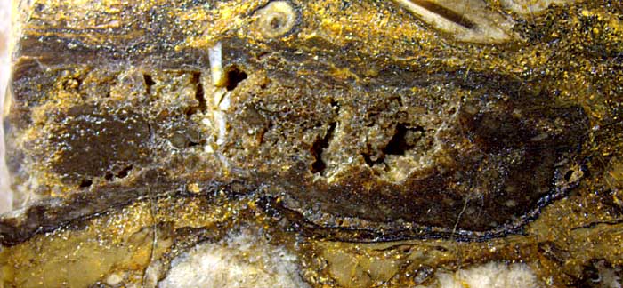
Fig.1:
Nematophyte, largely decayed and replaced by quartz grains and voids
inside,
filamentous structure partially preserved in some places near
the boundary. Width of the image 17mm.
With tube diameters ranging from well below 10µm up to 27µm and a width
of the whole organism of hardly
more than 2cm, this
nematophyte seems to be comparable to the flat one
shown
in Rhynie
Chert News 46
but it lacks the clearly structured outer sheath seen on that specimen.
In the
present state this nematophyte is bounded by a kind of envelope,
which had partially been detached before silicification. This
envelope possibly consists of dried-up organic gel, as suggested
by the
aspect of the better preserved nematophyte in Rhynie
Chert News 98.
Things
are slightly confusing since the organic gel between the filaments or
tubes becomes replaced by silica gel,
probably when the organic matter degrades and produces low pH as it is
known from degrading plants.
This specimen apparently got into the swamp matter and
remained there as a whole. (It is not seen here as a whole because it
slightly extends beyond the
edge of the chert sample seen on the left. 2cm-wide sections
of the whole specimen are on the back of this 5mm
slab and on the adjacent face of the next slab of 10mm, where the
specimen ends inside.)
Several
conclusions can be derived from the conspicuous former crack filled
with white chalzedony. It seems out of place among cavities left by the
decayed nematophyte weft. It
indicates that there had been some solid state, mechanically
homogeneous, probably silica gel from early silicification. (The
organic gel, which is thought to keep the live filaments together and
to keep the dirt out, would most probably not be so stiff that the weft
could break.) Bending stress on the weft gave rise to a crack starting
from the upper boundary and stopping at the lower boundary, which
indicates that the swamp matter outside was still fluid.
The silica-rich water in the crack turned into silica gel and
later into white
chalzedony. It remains an open question why the crack fill is still
there while the surrounding silica gel either vanished or
recrystallized into quartz.
A tolerably well preserved
weft of tubes with characteristic features is only seen near the right end in
Fig.1. The following pictures, width 1.4mm, show details from there.
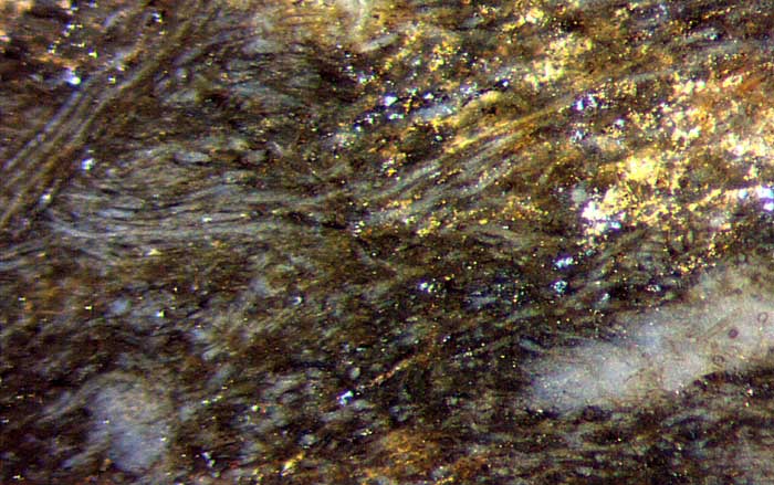
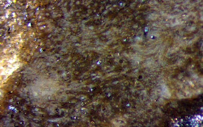
Figs.2,3:
Nematophyte tubes arranged not quite randomly: bundles of parallel or
diverging tubes, pale spots between the tangle of tubes.
Similar as with other nematophytes the tubes
seem to be more or less randomly arranged. The orientation can be governed by some
local texture, usually less conspicuous than the bunches in Fig.2. There
can be "glades
in the jungle", more or less clear spots of unknown
origin and purpose, as seen in the lower halves of Figs.2,3.
Their
abundance can exceed one per square millimeter. (The irregular bright specks indicate areas of the nematophyte
which had been decayed before silicification and hence are not relevant
here.) A diverging bundle of
tubes is poorly seen on the right in
Fig.3. A small part of the dark boundary is seen in Fig.3 below right.
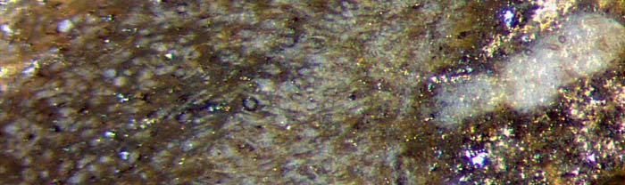
Fig.4: Nematophyte with narrow tubes near its boundary on the right, a
pale spot (incidentally ?) extending from there and
protruding into the surrounding chert with mineral debris.
Two of the
pale "glades" differ from the others by their elongated shape and
sparsely distributed narrow tubes of about 12µm, with the bigger tubes
keeping out. Their similar aspect seems to indicate that they
are no incidental formations but their purpose is not obvious. Also
seen in Fig.4 is an abundance of narrow tubes near an apparent boundary
of the nematophyte (incidentally ?) bordering on the pale
spot.
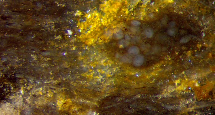
Fig.5:
Unidentified globules in a place of decayed and vanished nematophyte
tubes; degraded tubes on the left and below.
Globules like those in Fig.5, single or clustered, are seen scattered
across the whole nematophyte section. They have got a tiny dark rod
inside
which often appears as a black dot on the surface. Judging from their
deformation at mutual contact, they are of a soft consistence. Most
probably they do not belong to the nematophyte but prefer growing
inside it.
A large part of the weft of tubes in Fig.1, where not vanished at all,
looks as degraded as seen in Fig.5. A few thin dark lines seem to be
shrunken tube contents.
Any new find of a nematophyte, even if poorly preserved, can contribute
to an understanding of these rare "enigmatic organisms" [1].
H.-J. Weiss
2016,
revised Dec.2016, slightly modified 2017
[1] T.N. Taylor,
E.L.Taylor, M. Krings: Paleobotany, Elsevier 2009.
 |
 |
99 |