Scolecopteris
spores
Pinnules and sporangia of the "maggot fern" Scolecopteris
are common fossils in the Permian cherts of the Döhlen basin
but the related mature spores are seldom seen. More often one may find
immature spores within sporangia, possibly not yet full size.
Same
as with any other fern, the spores grow in globular groups of 4, called
tetrads, where they are symmetrically arranged such that every one
borders with 3 faces to neighbouring ones. The 3 plane faces of one
spore meet at 3 edges which meet at a common point where they make a
blunt tip. This can be seen after disintegration of the tetrad into
separate spores. The symmetry of the arrangement makes it resemble a
Mercedes "star". Hence, every spore has got a 3-fold symmetry axis.
"Stars"
with 3-fold symmetry are also seen at the 4 locations on the surface of
the tetrads where 3 spores meet. (Hence, one can expect to see "stars"
on randomy oriented tetrads 4 times oftener than on randomly oriented
spores.)
After the disintegration of the tetrads, the quarter spheres become
slightly inflated and more globular so that their blunt tip becomes
even blunter. With limited magnification it may be not quite easy to
distinguish between globular tetrads and globular spores. For
simplicity, all globular grains inside and outside sporangia are called
spores here.
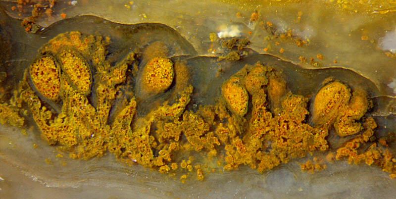
Fig.1 (above): Cross-sections of two neighbouring pinnules of the
maggot fern Scolecopteris
elegans
from Döhlen basin (Lower Permian), sporangia stuffed with yellow
spores, debris without relevant structure below. Width of the picture
4.3mm.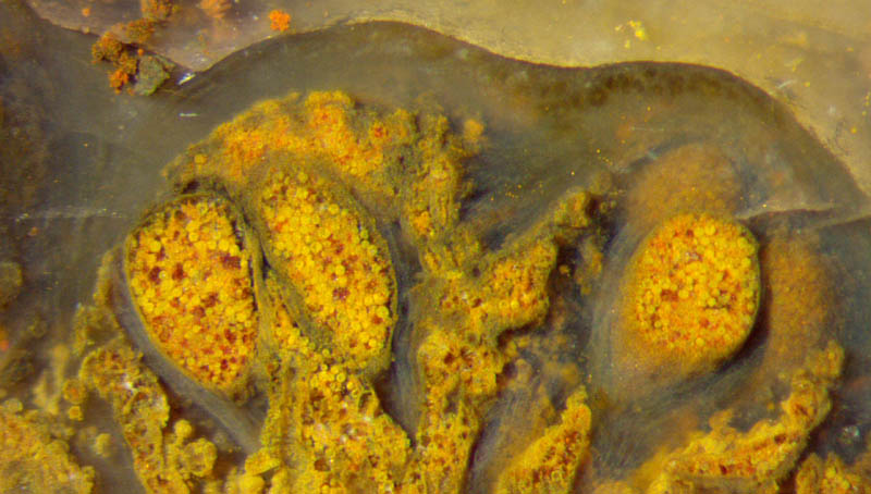
Fig.2 (right): Cross-sections of maggot
fern pinnule, detail of Fig.1. Width
of the picture 2.15mm.
The tissue structure of the pinnules and sporangia has largely
vanished so that the tissue is transparent now. On the right in Fig.2
one can see the fills of two sporangia through their walls.
The
big sporangium in Fig.1 on the right is cut open along a narrow strip.
On either side the yellow spores are seen through the wall.
Remains of the pinnule tissue are faintly seen in Fig.2.
As a conspicuous detail, the upper boundary of the pinnules is clearly
seen. This is due to the combined effect of biology, chemistry,
mechanics, and optics:
This fern, like all terrestrial plants,
protected itself against exsiccation by exuding substances to make a
thin cuticle on top of its epidermis. The cuticle is made of highly
rot-resistant organic polymers which persist through ages. The
interface between cuticle and chalzedony remains weakly bound. Under
mechanical stress, as by shrinkage, the interface provides an easy
crack path where a crack can start or an oncoming crack can be guided
along. If the crack is not much narrower than the wavelength
of
light, it reflects the incident light, which makes sthe sharp contrast
along the upper boundary of the pinnule.
Fig.3 (below): Longitudinal section of two sporangia of the
maggot fern, detail of Fig.1,2.
Width of the picture
0.8mm.
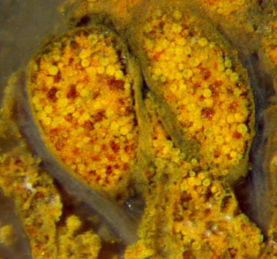
On Fig.3 one can see an opening in the sporangium on the
right, as it is known from Scolecopteris, with
some spores fallen out.
These sporangia are shown here in detail because the maggot fern from
Döhlen basin has been repeatedly reported in palaeobotany literature
with erroneous size data:
The sporangia in Fig.3 are shown in [1],
Bild 191, too large by a factor 1.7. (Fig.3 shows the present
state after polishing, hence there are small differences between the
pictures.)
The sporangium in Fig.2 on the right is shown in [2],
Abb.209, too large by a factor 2.7.
The sporangia in [3], S.70, are too
large by a factor 2.
The erroneous size data should have aroused suspicion even without
original samples available for comparison.
After [2], Abb.209, the spore size would have
been 70µm, which is bigger than the big spores
of Scolecopteris
macrospora
[4] and hence highly questionable. Checking against reality would have
provided their real size of hardly 30µm.
Contrary
to the too big size data of sporangia and spores in [1-3], other parts
of this plant are shown with size data too small by factors 3 to 10: [2],
Abb.210 und 130.
Erroneous size data are compiled under errors and
mistakes.
Sample: own collection, B/51.2 .
H.-J.
Weiss
2016
[1]
R. Rößler: Der
versteinerte Wald von Chemnitz. Museum f. Naturkunde Chemnitz, 2001.
[2] M.
Barthel: Die Rotliegendflora der Döhlen-Formation.
Geologica Saxonica 61 (2) 2015, 105-238.
[3] M.
Barthel:
Die Madensteine vom Windberg, in: U. Dernbach, W.D.
Tidwell (eds.):
Geheimnisse versteinerter Pflanzen, D'ORO 2002, 64-77.
[4] J.R..Jennings,
M.A. Millay: A new permineralized marattialean fern from
the Pennsylvanian. Palaeontology 21(1978), 709-716,
| |
 |
 15 15 |