Enigmatic cm-size blobs in Rhynie chert
Most samples of the famous Lower Devonian Rhynie chert show
silicified plant parts which are doubtless recognized as belonging to
one of the known early land plant species, often with lots of fungus
hyphae in between. Only a few samples contain distinctly preserved
algae, lichens, nematophytes, or cyanobacteria. More common are
indefinite layers and clouds probably indicating the presence of
microbes of unknown affiliation. Less common are clouds with more or
less well defined shapes or outlines, as the ones pictured here. It
may be worth trying to explain them.
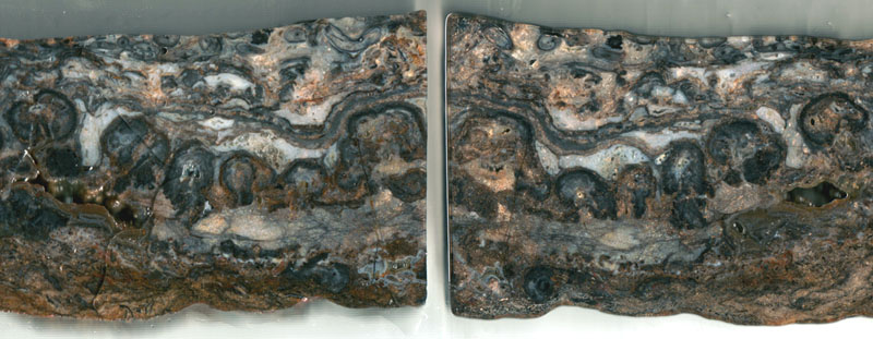 Fig.1: Two faces of one cut
through a layer fragment of Rhynie chert. Width of one picture 6cm.
(Figs.1-9: taken from vertical cuts of one
sample.)
With
a cut gap of about 2mm, and the two halves of Fig.1 not much differing
from mirror symmetry, it is obvious that the rounded shapes extend into
depth much farther than 2mm. As the two arrangements of the 6 blob-like
sections in Fig.1 are not seen on the rear of the 5mm slabs, it
can be concluded that they are sections of roughly globular
or slightly elongate blobs,
hence no remains of land plants and thus worth being looked at closely.
Their lower position in the chert layer indicates that they had been
there before Rhynia
grew or became flushed on top of them.
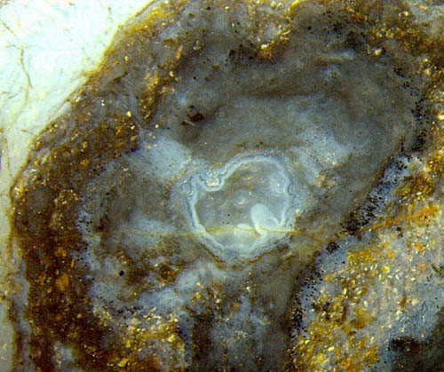 Fig.2 (left): One of the
apparent blobs in Fig.1 (left half, 2nd)
revealing a confusing structure: gray chalcedony with a former
cavity inside and surrounded with "glued" mineral debris. Width
of the picture 7mm.
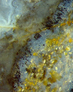
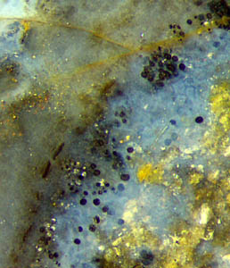
Figs.3,4:
Details of Fig.2, successively enlarged: globules
with dark or pale wall,
transparent (or white) inside, of various sizes
up to 35Ám, scattered along a strip of bluish chalzedony.
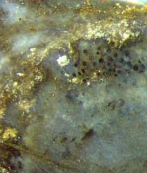
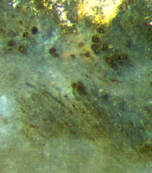
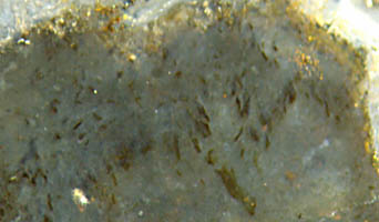
Figs.5,6 (below): Areas near the margin of the blobs with dark
globules
and indistinct bunches of dark streaks.
It
seems as if aligned filaments or tubes had been involved in the
formation of
the blobs but had mostly decayed and vanished from sight (Figs.5,6,7).
There are other peculiar features involved: black clots of various
shape, size, and arrangement. They are conspicuous when concave on the
margin as in Fig.8. The clots may be arranged along a boundary, at some
distance of it, or apparently at random. The tiny dark dots in
Fig.8 are quite different from the dark globules in Figs.2-6.
They seem to be the debris from a former structure. The
texture in Fig.7 is so
poorly preserved that it is hard to tell whether it originates
from tubes or from aligned (bunches of) filaments. Whatever it
was, it did not let the silt grains enter, hence they are kept
beyond the margin: Figs.2-8. (A similar
formation seen on another sample, with tubes
oriented towards the margin, has been discussed in connection with
nematophytes.)
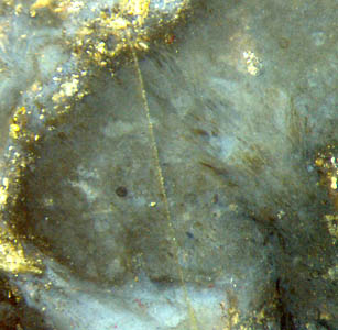
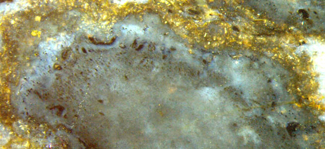
Fig.7
(far left): Area with a faint texture oriented towards the
margin. Width of the picture 3mm.
Fig.8: Upper part of a blob with light-coloured chalcedony
and dark substance of various shape. Width of the picture 6mm.
Fig.9 (below): Blob with black ribbon-like (?) remains of some
structure. Height of the picture 2mm.
 It seems difficult to make sense of these observations. At natural size
the blobs are seen with an apparently clear outline. Magnification
reveals that
there are several not so well defined
boundaries, more or less rugged ones.
What seems to be black globules in Figs.2-6
turns out to be hollow ones, now filled with clear chalzedony. If cut
into halves it is seen that their wall is not black but dark and
transparent. The black aspect against the background of light-coloured
chalzedony appears only if the light is
absorbed by both the front and rear wall of the whole
globule.
Apparently the wall is covered with tiny dark
dots about 6Ám apart. These
globules differ clearly from the chlamydospores
of the fungi whose hyphae are present in the
chert above the blobs in Fig.1.
The smallest globules tend to be not dark but
pale. A few globules in Figs.3,4 are of white aspect, which only
indicates that they became filled with fine-grained quartz.
Various tentative interpretations have to be
considered, including colonial microbes or algae, preferably arranged
in peripheral
positions around some of the blobs as if they formed a kind of
symbiosis. So it may turn
out that the small globules are no
characteristic feature of the cm-size blobs. Unfortunately, the
same can be suspected of the dark inclusions in Figs.5-9, too.
Without a practicable clue from the details, one can try to
derive a tentative interpretation of the blobs from the
overall aspect in
Fig.1: Roughly globular or half-spherical cm-size blobs attached to the
bottom under water or to moist soil are known from several
cyanobacteria
families. The blobs are usually kept in shape by organic
gel. Those considered here might have grown in clear water or on land,
then become flooded, with the silt particles sticking to their envelope
of gel where they later became silicified together with everything
else. Inclusions seen inside might either have been overgrown by the
cyanobacteria
or grown into the blob later,
then degraded. Large tubes found in this sample,
80...120Ám wide and thus being comparable to Palaeonitella,
may
look like the debris in Fig.9 when crushed.
The
attempted interpretations has to remain uncertain here because it is
not known whether the observed details belong together or have got together
on the images only incidentally.
H.-J.
Weiss
2014, modified 2015
 |
 |
68 |


 Fig.1: Two faces of one cut
through a layer fragment of Rhynie chert. Width of one picture 6cm.
 Fig.2 (left): One of the
apparent blobs in Fig.1 (left half, 2nd)
revealing a confusing structure: gray chalcedony with a former
cavity inside and surrounded with "glued" mineral debris. Width
of the picture 7mm.


 It seems difficult to make sense of these observations. At natural size
the blobs are seen with an apparently clear outline. Magnification
reveals that
there are several not so well defined
boundaries, more or less rugged ones.
