Cell-size clots in bennettitalean
tissue:
No oribatid mite coprolites
It is surprising to see how some dubious ideas
persist once they have
entered into the scientific literature. One notorious
example from palaeobotany concerns small dark clots in fossil plant
tissue for which a plausible explanation had to be found. As the clots
were usually seen in damaged parts of the tissue, the idea suggested
itself that they were droppings of some small creature feeding on the
tissue. This idea, believable as it
seemed, was fraught with more than one
problem: First, no fossil creature was seen near and far. This was
taken as an
excuse for introducing guesswork. Since mites are known to feed on
plant
matter, the mite coprolite idea was brought up. The guesswork became
even more specific: The mites, although elusive, were specified as
oribatid mites, and "oribatid mite coprolites"
became a popular term in the palaeobotany
literature since the 1990s. Those palaeobotanists
who readily adopted it had apparently not noticed an observational
fact: Where there are dark angular clots in damaged tissue, one can be
sure that their size and shape variation is compatible with the size
and shape variation of the cells of nearby intact tissue, which poses
another problem.
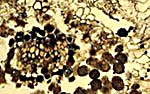
Figs.1,2:
Cell-size clots in damaged tissue of triassic
bennettitalean roots, details of Figs.6G,D in [1], interpreted
as oribatid mite coprolites there. Note the clot sizes fitting to the
largely differing cell sizes. In Fig.2 there are small cells filled
with clot matter in a coherent row, and larger loose clots on the right. Width
of the pictures 0.3mm (provided that the scale bars in [1] are correct).
It is hard to believe that it did not occur to those dealing with the
subject that such coincidence is strong evidence against the coprolite
interpretation. Perhaps it did occur later to those authors who
preferred not to propagate that interpretation any more. However, none
of them has expressly retracted the coprolite hypothesis. Therefore it
is necessary to comment on every such publication, which has been done
in the sequence as they have become known to the present writer. (See
Google: oribatid mite coprolites, or "Wood rot or
coprolites" on this website.)
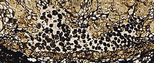
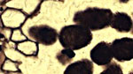
Fig.3: Cell-size clots in partially decayed tissue of a cretaceous
bennettitalean stem, detail of Fig.4E in [2], interpreted
as oribatid
mite coprolites there.
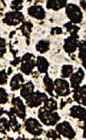
Fig.4 (right): Detail of Fig.3, polygonal outlines of angular clots
called
"spherical to ovoid" in [2].
The arguments concerning the misinterpretation of the clots are the
same as given repeatedly before. In case of good preservation, for
virtually every one of the loose clots an intact cell of fitting size
and shape can be found in the tissue, which strongly suggests the
conclusion that the clots had been some kind of cell casts which came
tumbling out when the cell walls broke down. Clot formation inside
cells and breakdown of the walls is most probably due to the same
cause. Fungi have been observed to form dense tangles of very thin
hyphae inside cells, and it is known that they are able to break down
cell walls of plants [3].
The clots in Figs.3,4 are described in [2] as "spherical to ovoid" with
"smooth
to slightly bumpy" surface but it is easily seen that the alleged bumpy
spheres are rather angular, often with polygonal outline, and even
right and acute angles are seen, as expected from replicas of the cell
lumen. This applies also to Fig.5.
The polyhedral clots usually misinterpreted as coprolites may be the
only fossil evidence for the original cell sizes and shapes of
vanished
or compressed tissues. The empty cells have retained their
shape in Figs.1,2 but part of them have become deformed or collapsed in
Figs.3,5.
Damaged plant tissue with the same aspect as in these images is usually
called
a frass gallery although there are no specific features of the damage
which would justify such interpretation. There are alleged galleries so
narrow that no herbivore could have crept there. There are alleged
coprolites in intact cells where no herbivore could have
deposited them.
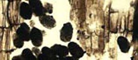
Fig.5: Angular clots in a partially damaged triassic
bennettitalean root, detail of Fig.6O in [1],
interpreted as oribatid
mite coprolites there.
Finally it can be concluded that the above evidence does not support
the hypothesis of oribatid mite activity in cretaceous and triassic
bennettitaleans. It remains to be checked whether or not other
reports on herbivore coprolites in bennettitalean fossils stand
a critical revision or have to be re-interpreted as wood rot as it has
been done with numerous cases of alleged oribatid mite coprolite
sightings in plant tissue.
H.-J.
Weiss 2013
[1] C.
Strullu-Derrien, S. McLoughlin, M. Philippe, A. Mørk, D.G. Strullu:
Arthropod interactions with
bennettitalean roots in a Triassic permineralized peat from Hopen,
Svalbard.
Palaeogeography, Palaeoclimatology,
Palaeoecology 348–349(2012), 45-58.
[2]
N.A. Jud, G.W. Rothwell, R.A. Stockey:
Paleoecological and phylogenetic implications of Saxicaulis
meckertii ... :
A bennettitalean stem from the Upper
Cretaceous ...
Int J. Plant Sci. 171(2010), 915-25.
[3] T.N. Taylor
et al.: Paleobotany. Elsevier 2009
|

|
 18 18 |