Well-known creatures and enigmatic
formations in one piece of chert
Fossil
remains of early land plants, microbes, and fungi are abundantly
preserved in the Rhynie chert. Much less abundant but not really rare
are little creatures and their moults. Really rare are samples with
more than one species of creatures inside, like the sample presented
here.
Most easily recognized are the cross-sections of trigonotarbids
and their exuviae shed in moulting, often with hairy legs nearby. The
slight inclination of the exuviae
with respect to the cut plane in Fig.1 can
give the impression of a thick wall but this is a mere illusion since
the
wall is very thin. Legs are clearly seen
as cross-sections on the
left. Legs are essential body parts for those spider-like predators
with
solitary
ways of life on dry land.
Quite different is the aspect of the outwardly
legless freshwater
crustacean Ebullitiocaris
(Fig.2): This egg-shaped, apparently mainly sessile creature feeding on
algae is usually seen with
several specimens near
each other, and occasionally even in large numbers closely spaced and
clinging to inundated terrestrial plants.
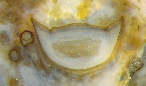
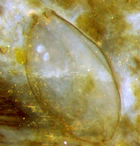
Fig.1 (far left): Trigonotarbid
(Palaeocharinus) exuviae
of body and two legs shed in moulting, thin-walled, slightly tilted
cross-sections, with bluish chalcedony deposited inside along the wall.
Fig.2:Ebullitiocaris,
egg-shaped enclosure with an
opening above as the only distinctly
seen detail of the body. Height of Figs.1,2: 1mm.
This chert sample seems to be the only one
offering both Palaeo- charinus
and Ebullitiocaris.
In addition to the creatures easily recog- nized as such, it offers three
phenomena with had been described repeatedly but not fully explained. Probably they are seen here
for
the first time combined in one plant cross-section: Fig.3.
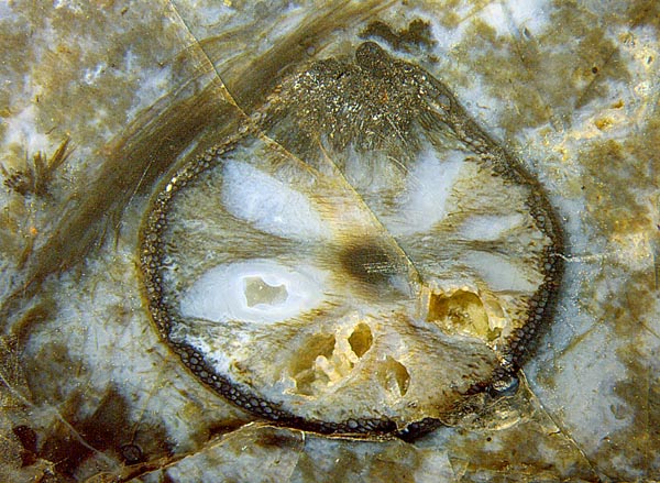
Fig.3 (right): Cross-section of Aglaophyton thrice
affected, (image width 8mm):
(1) enigmatic "flower-shaped" void pattern
in the tissue,
(2) tissue
decayed except for a peripheral layer of cells: "hollow straw aspect",
(3) large scab due to misguided growth.
ad (1): Judging from other specimens (see
Rhynie
Chert
News 4), the
cavities
seen radially arranged on
cross-sections are extending along the shoot, thereby
even passing
forking sites. Their occasional occurrence on some of the Aglaophyton
specimens remains enigmatic.
Most probably, the cavities in
the upright living plant had been empty.
ad (2): After formation of the void
pattern most of the tissue in between decayed except
for surprisingly well preserved cells along the
circumference which probably had been able to keep the shoot alive.
ad
(3): At some early stage the plant had developed a growth anomaly
causing the tissue to grow a large scab, locally interfering with the
void pattern
and "hollow straw" formation.
The phenomena described so far could well have been there in the
upright
living plant
while trigonotarbids prowled around. Eventually the vegetation
became inundated in flooding. Microbes and algae thrived, providing
food for the crustaceans dwelling between the plant remains. All
cavities filled with water, included those which are empty now. For
reasons
unknown, chalcedony deposited itself differentially in the water-filled
cavities inside the cross-section in Fig.3. While the cavities below
did not get filled with
chalcedony, some aquatic
fungus hyphae got the opportunity
to grow there in the water. Later they became coated with tiny quartz
crystals. After the supply of silica by diffusion
had stopped, the cavities remained empty
as they are now, crossed by coated hyphae.
The
cuticle on the surface of the plant reveals its presence by deflecting
the crack approaching from below left in Fig.3 such that it follows the
plant
surface until it runs into a crushed area.
Also worth mentioning
here is a fact already
discussed in Rhynie
Chert News 105:
The wall thickness of the "hollow
straw" does not
represent the silica diffusion depth as claimed in [1] but is brought about by
the ability of the
living plant to protect a fraction of the tissue against decay. Surprising evidence is provided by a very seldom seen phenomenon, shown in Rhynie
Chert News 60,
there Fig.4: At an early stage, local damage at the periphery had been covered with a cap whose cells, like those along the periphery,
had been made decay-resistant in the live plant by unknown means.
Hence, the wall thickness of the hollow straw, like that of the cap,
had been brought about by processes in the live plant.
As noticed as early as 2010 and mentioned anew in 2014 [2], Ebullitiocaris
is erroneously called rotifer in "Paleobotany" [3]. (See Errors and
Mistakes. This erroneous interpretation has been included into
fossilhunters.xyz ebullitiocaris:
... Peel Technique, Figure 157.)
Sample: Rh2/176.2, obtained from Shanks
in 2012.
H.-J.
Weiss
2021
[1] www.abdn.ac.uk/rhynie
[2]
H.-J.
Weiss: Rhynie
chert – Implications of new finds. EPPC 2014,
Padua.
[3]
T.N. Taylor,
E.L. Taylor, M. Krings: Paleobotany. Elsevier 2009.
 |
 |
179 |