Glades in the nematophyte jungle
Nematophytes are still enigmatic as a whole and in detail as
well.
One detail, lately seen on a cut face of a Rhynie chert sample with the
nematophyte Nematoplexus,
incidentally illuminated by the reflected light from a crack and thus
visible in Fig.2 of Rhynie
Chert
News
152, has
led to
thorough redrafting of this contribution. There
one can see how one of the spiralling Nematoplexus
tubes begins without branching at a position which must be at the
boundary of a central clot. Hence, the widespread view of the "branch-knots"
of Nematoplexus
as
a tangle
of tubes, with "tubes
... entering and leaving" [1], is not true.
(Without
branching and without
"entering and leaving" tubes,
the term "branch-knot"
is misleading and should be avoided.)
A comparison of the big clots of this unknown
nematophyte, appearing
as "glades" in cross-sections, with the small clots in the tangles of Nematoplexus suggests
itself. The big clots, too, are not traversed by tubes, and the tubes
begin possibly at
or near the surface. Apart from the similarities there are obvious
differences: These tubes are much bigger, and they are nearly parallel.
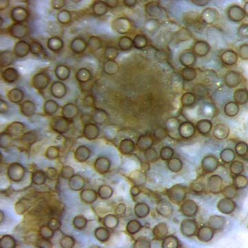
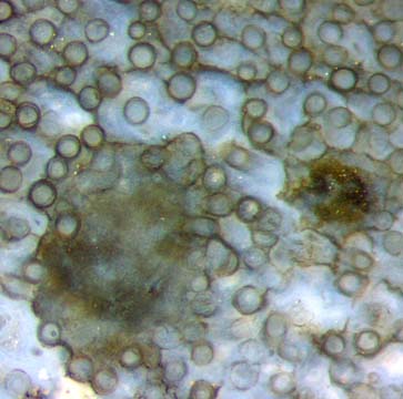
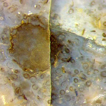
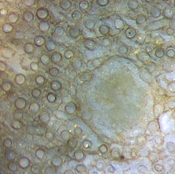
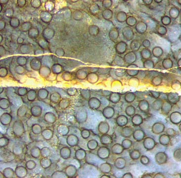
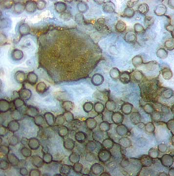
Figs.
1-7: Nematophyte
consisting of rather well aligned tubes,
mainly
50-60µm across,
randomly distributed between
sections
of rounded lumps seen as
"glades"
of 0.35-0.5mm
with a
frequency of
about 10/cm2.
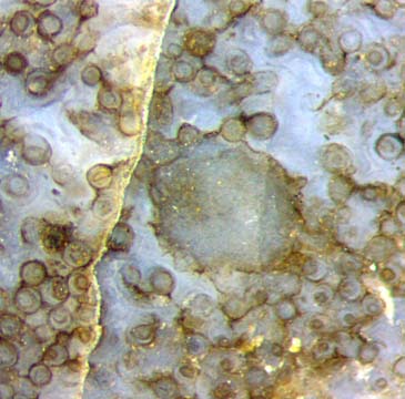
Fig.3 (in the upper row):
Area with tubes of 15-60µm width.
The size of every image is 1mm2.
There is no good lengthwise cut of this nematophyte
available at present so that evidence of
tubes emerging from the clots is poor. A few short tube parts with
deviating
directions are seen in Fig.2. One or two deviating tubes in
Fig.6 seem to emerge
from the surface of the clot (above right). In Fig.7, one tube fits
perfectly to the periphery of the clot (above).
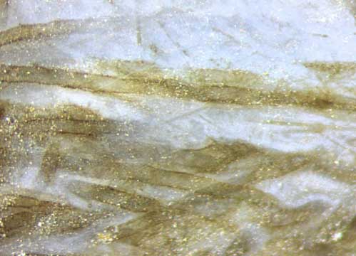
Fig.8:
Nematophyte tubes, slightly deranged and separated, thus individually
visible in lateral view.
The tiny white dots
are due to
the roughness of the raw sample
surface. Same sample and scale as above.
Judging from small fracture faces along the
densely spaced
tubes which offer a sideways view, even
a polished
longitudinal section would only offer a
confusing assembly of lines with poor contrast which would
not look like tubes. Individual tubes can be
seen in lateral view if they are displaced
and separated by white chalcedony as in Fig.8.
The small-scale waviness
of the tube walls in Fig.8 seems to indicate that the tubes had been
rather soft
before silicification, as also observed
in Rhynie
Chert
News 40.
The stronger contrast in Figs. 1-7
compared to Fig.8 does
not indicate better
preservation or thick-walled tubes
but
is simply an optical effect. Looking in tube direction in transparent chalcedony
makes a sharper contour
than looking across. A thin microbial layer would enhance this effect.
Other microbial sheets
are seen as black lines connecting the tube sections, as in Fig.6.
Annotation 2020: Thanks
are due to Gerd Schmahl for
microphotographs, particularly
for
Fig.2 of Rhynie
Chert
News
152, which
has suggested a re-interpretation of the above pictures.
With the assumption that the clearly
seen "glades
in the nematophyte jungle"
are comparable with the less well seen "knots" or "medullary spots" of
other nematophytes, one may compare the unknown nematophyte
with Nematoplexus
and Prototaxites.
By doing so, one may conclude
that the knots of Nematoplexus
have got a definite boundary although it is usually not seen. Also one
may guess that the tubes of the above nematophyte and others
emerge from the periphery of clots seen here as "glades", also known
as "medullary
spots".
The much disputed Prototaxites,
once
regarded as an enormously big nematophyte
but lately as a huge fungus or lichen [2,3], raises the question
whether or
not other nematophytes are rather fungi or lichens. Various
nematophytes, including Nematasketum and
tubes with sizes like those in Figs.1-8, are presented in [4] and
commented on: "However,
they are twice the diameter as the skeletal tubes in Nematasketum
(Burgess and Edwards,1988)" [4].
It is not attempted here to assign the nematophyte in Figs.1-8 to one
of the already described species.
Sample: Rh2/81,
0.63kg, obtained from Shanks in
2003, pictures taken from Part
3.
H.-J.
Weiss 2016,
revised in 2020
[1] A.G. Lyon:
On the fragmentary remains of an organism referable to the
nematophytales, from the Rhynie chert, Nematoplexus rhyniensis.
Trans. Roy. Soc. Edinburgh
65(1961-62), 79-87, 2 tables.
[2]
T.N.Taylor,
M. Krings, E.L. Taylor: Fossil
Fungi. Elsevier 2015.
[3]
R. Honegger, D. Edwards, L. Axe, Ch. Strullu-Derrien:
Fertile Prototaxites taiti: a basal ascomycete with
inoperculate, polysporous asci lacking croziers.
Phil.
Trans. Roy. Soc. B 373 (2017): 20170146.
[4] P. Filipiak, H. Szaniawski:
Nematophytes
from the Lower Devonian of Podolia, Ukraine.
Rev. Pal. Palyn. 224 (2016),
109-120.
 |
 |
92 |