Degradation and preservation of
palaeozoic wood
Petrified wood is known in various states of preservation. It
may reveal tiny details of the tissue or consecutive stages of decay
before and during silicification, also damage to the living tree. A few
related observations are reported in the following. As
a remarkable
fact, severely decayed wood is often seen immediately beside well
preserved tissue on cut faces of silicified wood (Fig.1). This
suggests the idea that highly selective processes had been at work
which affected individual cells, often spreading from one cell to the
next, leaving some cells well preserved but others completely decayed.
The pictured sample is problematic as there is more than one
thinkable explanation of Fig.1. The distorted pith rays above seem to
indicate that first came a process spreading preferably along the
radial cell files and pith rays, filling the cells with transparent
chalcedony with a brown hue, which makes a dark aspect if one looks
into the tracheid tubes. The cells preserved in this way were not
accessible to the subsequently encroaching rot which caused disorder by
weakening or consuming the cell walls of larger parts of the wood,
giving
rise to the deposition of whitish chalcedony and to the growth of crystalline quartz
so that a few distorted pith rays is all that is left of the former
structure.
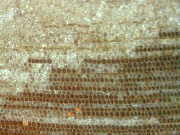
Fig.1: Well preserved radial cell files adjacent
to decaying coniferous-type wood indicated by a few distorted pith rays
above. Cell diameter about
50µm.
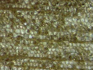 Another
sign of selectivity is seen in Fig.2, where individual cells are
(partially) filled with one dark clot each, which resembles the clots
produced by fungi in extant wood [1].
Another
sign of selectivity is seen in Fig.2, where individual cells are
(partially) filled with one dark clot each, which resembles the clots
produced by fungi in extant wood [1].
Fig.2, right: Dark clots, often smaller than the cells, scattered
throughout the tissue. Same sample as Fig.1.


Figs.3,4: Clots within
cells in radial files.
A combination of
the two features making rounded dark clots within cells in radial files
has been
observed in Permian wood from Germany (Fig.3) and China (Fig.4).
The
latter clots have been questionably interpreted as oribatid mite
coprolites [2].
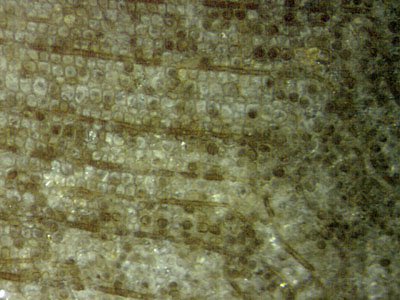 Fig.5 (right): Intact wood structure
on the left bordering on disintegrated tissue on the right. Note the
dark
pith rays.
Fig.5 (right): Intact wood structure
on the left bordering on disintegrated tissue on the right. Note the
dark
pith rays.
Degradation
or loss of strength may reveal itself by large irreversible deformation
under load as seen in Fig.5, where there is a
rather sharp boundary
between the regular wood structure on the left
and the distorted tissue on the right. Although the
contributions
of the
various processes possibly involved in degradation are not known, it
can be stated that
large shear deformation or kinking is usually not the cause of decay,
as it is also seen in this sample where one can find small internal
kinks
without disintegration (Fig.6). Hence, Fig.5 suggests
that first came local degradation, then local shear and decay.
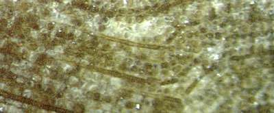 Fig.6 (left): Kink in wood without
disintegration of
tissue.
Fig.6 (left): Kink in wood without
disintegration of
tissue.
Obviouly
the cells had not been empty during deformation, otherwise
they
would have been squeezed into rhomboid or oblique shapes as often seen
in petrified wood, also in Fig.7, below left.
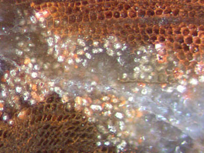
Fig.7: Light-coloured clots representing cell
lumina of
disintegrated wood next to dark areas of coherent tissue stained red
with hematite. Width of the picture 1.2mm.
Loss of mechanical strength of the wood,
revealing itself as large deformations, must be
due to weakening of the cell walls. This can be inferred from Fig.5
but is not immediately obvious.
It
is obvious from a peculiar variety of local disintegration as in Fig.7,
where
the cell walls were essentially consumed, and if the cell lumina had
not
been preserved somehow as white clots, one would not have a strong
argument for the assumption that there
had been wood
with cells in radial files where chaos reigns now. Incidentally, this
image is different since
there is a horizontal dark line which mediates between the random clot
distribution and the regular tissue.
Larger random assemblages of whitish clots
had been mentioned earlier [3] and will be the subject of a forthcoming
contribution. If not adjoining to
well-preserved tissue as in Fig.7, the clots may easily be mistaken for
remains of the central pith. Clots of this kind, like the more
frequently seen dark ones, have repeatedly been mistaken for mite
coprolites.
The samples in Figs.1,2,5,6,7 were found at the
Borxleben gravel pit near the Kyffhäuser Mountains,
Germany, uppermost Carboniferous [4]. The sample in Fig.7 has
been provided by W.+G.
Etzrodt, Borxleben, who also supported the collection of
other samples at the location. The
sample in Fig.3 (Lower Permian)
has been provided by Ch.
Krüger , Schallodenbach, .
Sample labels: Figs.1,2,5: KyB/112.1, Fig.6:
KyB/112.2,
Fig.7: KyB/12.1,
The observations are compatible with the following
interpretation:
- There had been interrelations between processes promoting
decay or preservation.
- The processes did not work simultaneously throughout but
locally by spreading of affected areas, resembling the spread of rot
in live or dead wood.
- The loss of
strength and
decay of cell walls had probably been
brought about by fungus activity.
-
Metabolic waste released by some fungus attacking the wood and /or
substances from partially decaying wood triggered the precipitation of
SiO2 , with a
tendency to preserve what had
been left of the tissue.
- The processes of both decay and preservation could
have begun with
individual affected cells and subsequently spread across the tissue.
-
The rich non-biological structure of fossil wood from
several
locations, notably of the type found at the Kyffhäuser mountains, is
the result of competing processes whose unravelling has just begun and
will reqire much more effort.
Discussing the formation of
non-biological structures mediated by biological agents is not idle
talk since the subject has been the source of misinterpretations
by
professionals from about 200 years ago until present.
H.-J. Weiss
2013
[1] F.
Schwarze: Fungal strategies of wood decay in trees.
Springer, Berlin 2004.
[2] Zhuo
Feng, Jun Wang, Lu-Yun Liu:
First report of oribatid mite (arthropod) borings and coprolites in
Permian woods from the Helan Mountains of northern China.
Palaeogeography,
Palaeoclimatology, Palaeoecology 288(2010), 54-61.
[3] H.-J. Weiss:
Beobachtungen an Kieselhölzern des Kyffhäuser-Gebirges.
Veröff. Mus.
Naturkunde Chemnitz 21(1998), 37-48.
[4] J. Schneider,
R. Rößler,
B. Gaitsch: Stratigraphy and facies of the Middle
European continental Carboniferous and Permian excursion guide A5, 1995.
|

|
 21 21 |