On the origin of white spots in fossil
wood
The
usually dark aspect of thoroughly silicified wood is brought about by
the transparent chalcedony or quartz which lets the light penetrate
into depth where it is trapped among cell walls and decay products of
the wood. The presence of white spots indicates that some process had
been going on which deviated from the usual way of silicification.
Since
the phenomenon is found in silicified wood from several locations and
may be quite common, it has aroused the interest of palaeobotanists and
fossil collectors, which apparently did not lead to an explanation. The
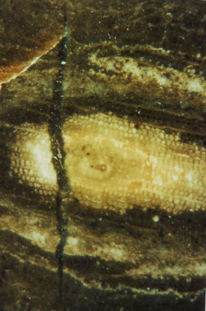
spots are often structured (Fig.1) and occasionally shaped, sized, and
arranged on the tree trunk cross-section such that the whole may
resemble some poorly preserved Psaronius stem, and really had been
repeatedly mistaken for such, once even by the famous B. Cotta
[1]. 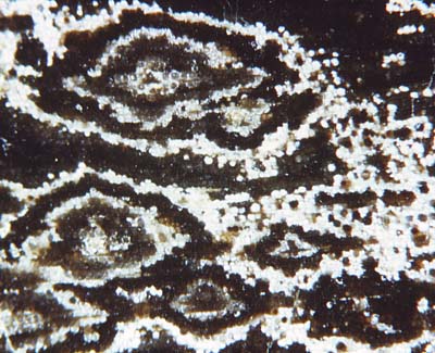
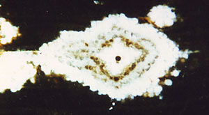
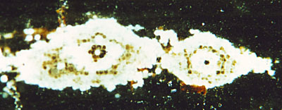
Figs.1-3:
Fossil wood cross-section with structured spots due to some peculiarity
of the silicification process which affects individual cells. (The
radial direction is horizontal. Note the bloated cell in the centre of the spots in Figs.2,3.)
The
aspect suggests the notion of growth beginning at a centre and
proceeding with some enigmatic periodicity. (See also "Silicified wood".)
The centre of growth is
distinctly seen in every structured spot
in Figs.2,3 as one cell cross-section
bloated into circular shape. As
a most peculiar fact, the central cell of the white spot can be either
dark or white. Hence, one may well suspect that the explanation of the
whole phenomenon must be rather complex. No explanation will be
proposed here but details are described which eventually will lead the
way.
The straight border on the left in Fig.2 is due to a
pith ray which traverses the big spot and is vaguely seen coming out of
the spot on the right.
The centre may consist of more than one cell, as seen in Fig.4.
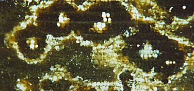
Fig.4: Fossil wood cross-section, cells with
white fill partially arranged along radial tracheid files.
Here, a tendency of the process to propagate along the radial cell
files of the wood is obvious, in addition to the other tendency to form
concentric zones. Not visible on these cross-sections but worth
mentioning is the strongest tendency, which is propagation along the
tracheids, so that in 3D, the spots are really slender rods.
It had been proposed that these Upper Carboniferous wood specimens developed
the spots much later, possibly in the Tertiary, by a similar process
which bleaches flint while exposed to the sun [2]. That idea can be
refuted
by several observations indicating that the spots were early
formations [3].
The
short crack in Fig.5, whose gap is rather wide considering that its
length does not exceed the picture frame, must have formed
while
the whole was still soft, a silica gel in an intermediate state of
solidification. Obviously the white spot is older than the crack, hence
was formed early. From the fact that the wood structure is well
preserved in the spot but largely deformed and decayed in the vicinity
it is evident that the spots are areas of early mineralisation which
prevented decay, and that
some time must have elapsed between spot formation and solidification
of the whole.
Fig.5: Fossil wood cross-section with a
structured spot transversed by a crack indicating a
gel-like consitution of the whole while the crack was formed.
It
can be hoped that the observed details reported here, if combined with
further investigations, will lead to a satisfactory explanation of the
phenomenon.
Samples found among gravel near Kyffhäuser Mountains,
Germany, uppermost Carboniferous,
provided by W.+G.
Etzrodt,
Borxleben. Photographs by
R.
Noll, Tiefenthal.
Sample labels: Fig.2: KyB/29.2,
Fig.3: KyB/29.3, Fig.5: KyB/6.1
H.-J.
Weiss
2011
Annotation 2012: Recently a beautifully patterned
coniferous-type fossil wood sample has been misidentified as Tempskya
by H. Kerp.
See Mock
Tempskya.
[1] B.
Cotta: Die
Dendrolithen in Bezug auf ihren inneren Bau. Leipzig und Dresden, 1832.
[2] H. Süss, P.
Rangnow: Die Fossiliensammlung Heinrich Cottas im
Museum für Naturkunde Berlin, Neue Museumskunde 27(1984), 17-30.
[3] H.-J.
Weiss: Veröff. Mus. Naturk. Chemnitz 21(1998),
37-48.
|

|
 2 2 |

 2
2


 2
2