Psaronius
conducting
strands with peculiar aspects
The tracheids
making up the conducting strands in the centre and in the aerial roots
of Psaronius
tree ferns are known to show scalariform wall patterns if
suitably preserved, as in Fig.1. There, a crack had taken a path along
two tracheid walls, seen right and left of a tracheid with bluish fill
showing a faint
opalescence. The picture plane has been chosen such that it essentially
coincides with the crack face on the natural surface of this chert
sample.
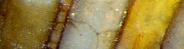
Fig.1: Two
Psaronius
tracheid walls on the raw surface of a natural chert
fragment, with "scalariform" pattern, steps
10µm. Width of the picture 0.87mm.
Psaronius
tracheid walls
of quite different aspect are seen in another chert sample in Fig.2.
They appear as an arrangement of
surprisingly straight and uniform thin planks, resembling an artistic
design rather than plant remains.
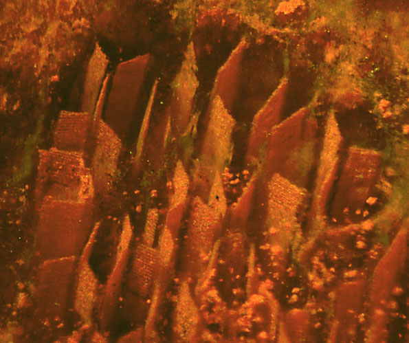
Fig.2 (left): Psaronius tracheid
walls
seen here as if emerging from the depth below the
cut face of the transparent
chalcedony.
Width of the picture 1mm.
With some effort one may see the scalariform pattern
on a few walls in Fig.2, as in the lower left quarter of the
picture. Owing to both foreshortening and slightly
lower
magnification, the patterned walls are less
well visible than those on
Fig.1.
The triple points or lines where 3 tracheid walls meet
are prone to decomposition before silicification so that the
tissue appears disintegrated into a bundle of straight walls here. The
thickness of some of them seems to be as low as 3µm or less. Hence, the
visible pattern must be only an imprint
of the scalariform structure seen in Fig.3,
after the larger part had broken off.
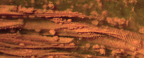
Fig.3: Psaronius
tracheid walls with larger parts of the scalariform structure still
adhering, seen at the cut face and below
in transparent chalcedony. Spacing 10-11µm.
Width of the picture 1mm.
Same scale, same sample as Fig.2.
In
this sample, most conducting strands with well visible tracheid walls
similar as in Fig.2 show a peculiar feature: The tracheids positioned
around the cirumference of the bundle look like empty thin-walled
boxes with one or two walls missing. There are more of them seen as
cross-sections in Fig.4. The
place where the outer wall must have been can be guessed from the
colouring in some cases.
Why the outer
walls of the outer tracheids are missing while the inner walls are
conspicuously distinctive, remains an open question here. There may be
no walls missing on conducting strands in other
Psaronius
samples, as in Permian Chert
News 12, for example.
The tracheids are of various shapes: Their
cross-sections vary between triangle (below) and septagon (above
right). The walls appear much thicker than those in Fig.2. (Note the
factor 2 in magnification.)
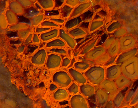
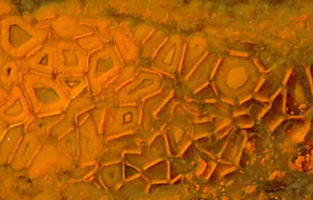
Fig.4: Psaronius
conducting strand cross-section with apparently separate or missing
tracheid walls. Width of the picture 1.5mm.
Fig5 (far right): Psaronius
conducting strand cross-section with cohering tracheid walls.
Width of the picture 1.5mm.
There is a conducting strand
of less-common aspect in the same sample:
Fig.5. Here,
either of two adjoining tracheids contributes to
the common wall apparently consisting
of three layers, with the thin dark middle layer
probably representing the 3µm-walls as seen in Fig.2. Considering that
the middle layer had been formed from two cell walls, it is a double
layer, although not visible as such in the present images.
Apparently the composite walls can
disintegrate,
beginning at the triple points of the network.
Similar thin lines along the walls between tracheids
of conducting
strands have also been seen on other Psaronius
cross-sections, though less distinct: Permian Chert
News
12
, (there Fig.6).
The thickness of the composite walls in Fig.5, 20...25µm,
is
about the same as that of the walls in Fig.3 with
a row of tubes on either side, as expected. The wall pattern elements,
seen here as straight tubes, are usually thought to be a
means of stiffening, which seems questionable.
Small pale spots on part of the raw outside
of this sample, which is deep red inside throughout, offer a
surprisingly different aspect of the conducting
strands: Fig.6. Apparently the chalzedony had recrystallized in a thin
surface layer of
this trunk fragment and thereby turned whitish, as it is known from
flintstone,
for example. Other than the transparent chalcedony in the above
pictures, the whitish
aspect is due to light reflection at crystals whose sizes exceed the
wavelength of light. The coarser crystalline structure also reveals
itself by slowly soaking up water, thereby turning dark.
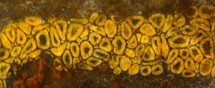
Fig.6: Psaronius
conducting strand broken across, of different aspect resulting from recrystallization at the surface, same trunk
fragment as the polished cut faces in Figs.2-5. Width of the picture
2.5mm, same magnification as Figs.4,5.
The
tracheid fills, conspicuous in Fig.6, less so in Figs.4,5, and nothing
except clear chalzedony in Fig.2, seem to be the result of locally
differing
conditions during silicification. The largely different aspects as
represented
by Figs.2,4,5,6 may be found even within one conducting strand, with
smooth
transitions between them. They may also be found in the central strands
of the aerial roots. (Related pictures will be shown in a forthcoming
contribution.)
As
mentioned above, the colour of Fig.6 is an exception since it has
formed after fragmentation of the petrified Psaronius trunk,
which
could have been as late as in the Cretaceous, judging from tiny remains
of
sandstone sticking tightly to the surface of the trunk and to the old
fracture faces.
This sample is a fragment of a Psaronius
tree trunk apparently
collapsed from circular into flat cross section when the parenchyma
between the conducting strands decayed. Apparently there had not
been more squeezing involved since the conducting strands and
their
cells do
not look squeezed. Often the centre with the conducting strands is
more squeezed, as in
Permian
Chert News 6.
This sample is one from the rare "wine-red" variety of the
"red", or rather ocre,
fossiliferous cherts in the Lower Permian Doehlen basin in Saxony,
Germany. It had been found by
S. Weiss in about 1991 as
the only piece
of red chert in a glacial stream deposit cast open on the area of the
Wilmsdorf golf course near Dresden.
The samples are kept in the own collection under the
labels W/19 (Fig.1) and W/20 (Figs.2-6).
H.-J.
Weiss 2019
|
 |
 25 25 |