A well preserved Psaronius fragment
from
Döhlen Basin
Stems of the tree fern Psaronius
are not rare in the fossiliferous cherts of the Döhlen Basin. They had
been silicified along with fragments of the related foliage while lying
prostrate in a swamp. Usually they were in a state of partial decay and
squeezed flat before accumulating enough silica gel for final
stabilisation of the shape. Even with the best preserved stem fragment
found so far, only the aerial roots have retained their shape but the
centre of the stem, where the stabilizing silica got latest, was not
able to withstand the pressure so that most of
the xylem and sclerenchyma
strands collapsed. (See Permian
Chert News 6.)
So the unexpected
find of a fragment of an uncommonly well preserved central part of
Psaronius remains
a cause of wonder (Fig.1).
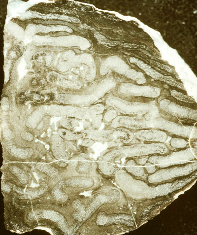
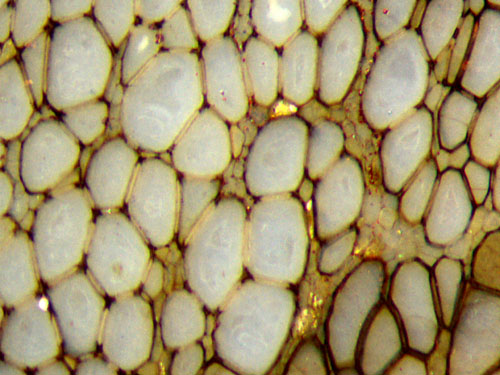
Fig.1: Cross-section of a (half) Psaronius centre,
well preserved, locally
deformed, estimated original diameter about 10cm, width of the picture
4cm. Photograph: M. Barthel.
With
its dark aspect and the virtual absence of any colour it differs
conspicuously from all specimens found in the chert but rather
resembles the abundant fragments of fossil wood. Possibly it
has never been
a constituent of a chert layer but became silicified along with
coniferous-type tree trunks while embedded in volcanic "ash".
Most
of the conducting strands with scalariform tracheids have
retained their original shape while the parenchyma between the strands
collapsed under slight compression in some places. As a result, the
strand cross-sections seem to converge to the
right. What is most
interesting about this sample is not its overall aspect seen in Fig.1
but the details of the well preserved tissues.
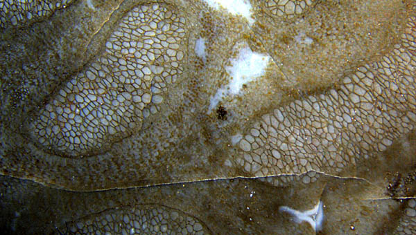
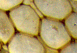
Fig.2 (left): Psaronius
conducting strands embedded in slightly damaged
parenchyma. Width of the picture 11mm.
Fig.3
(below): Several phenomena revealed by magnification of Fig.2: small
cells supposed to be phloem next to the big xylem
tracheids,
a small sclerenchyma strand stained black, parenchyma locally
replaced by
cell-size clots.
Width of the picture 3.6mm.
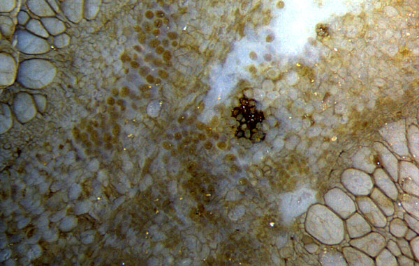
As seen in Figs.2,3, there are places on the cross-section where the
tissues are apparently not deformed.
The small
cells near and between the xylem
tracheids, supposed
to be phloem, are not clearly separated from the larger parenchyma
cells.
By comparison with numerous similar phenomena it can be stated
that the dark clots
in places of damaged tissue (Fig.3), usually
thought to be mite coprolites, also in the case of Psaronius [1], are
the result of fungus activity inside cells.
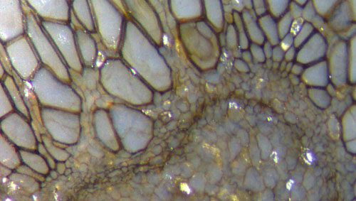
Figs.4,5,6 (left and below): Details of Psaronius
conducting strands.
Width of the pictures 1.4mm, 0.44mm (Fig.6).
The images show several features which seem to be
characteristic for the conducting strands of Psaronius:
(1) The tracheid cross-sections differ largely in shape and size.
(2)
At some spots on the outside of the conducting strands, tracheids
seem to vary from phloem size to full size (Fig.4).
 (3) The aspect of
cross-sections strongly suggests the presence of a
gap of 10-15µm between adjacent tracheids
(Figs.4,5), which turns out to be an illusion due to the absence of a
visible middle lamella in most cases.
(3) The aspect of
cross-sections strongly suggests the presence of a
gap of 10-15µm between adjacent tracheids
(Figs.4,5), which turns out to be an illusion due to the absence of a
visible middle lamella in most cases.
(4) A middle lamella
is seldom seen as a very thin dark line along the apparent gap (Fig.6).
(5)
The dark aspect of the tracheid walls (Figs.2-5) is a secondary
phenomenon. The deposition of a dark stain begins at the junctions
between the tracheids and may spread along the
colourless cell walls (Fig.5).
(6)
Phloem cells are distributed among the tracheids such that nearly every
tracheid cross-section is seen to contact one or more phloem cells
(Figs.3-6). Hence it can be concluded that every
one of the long and wide tracheids contacts many small phloem cells.
(7) The tracheid walls are covered with a "scalariform" pattern (Fig.7).
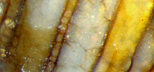
Fig.7 (right): Psaronius
tracheid walls on a fracture face at the
lateral surface of the
sample, two of them revealing a faintly seen "scalariform"
pattern with steps of 10µm. Note also the faint opalescence in the middle. Width of the
picture 1mm.
A few complementary remarks may be appropriate here.
- The tracheids of Psaronius
have often been pictured as components of the well-known "star-shaped"
cross-sections of the conducting strand in the aerial roots
but rarely in connection with the conducting strands
in the stem centre as in [2], Plate 35.
- The middle lamella along the
apparent gaps between tracheids as faintly seen in Fig.6
is also pictured in [2] (The wide gap in Fig.6 above consists of one or
two narrow phloem cells.)
-
The pockets of small tracheids as in Fig.4, irregularly distributed at
the boundary of the conducting strands, seem to be analogous to the
small tracheids at the tips of the
"star-shaped"
strands in the aerial roots [2].
-
Cracks in the chalcedony fill of the tracheids as seen in Figs.3,4,7
are liable to be mistaken for remains of cell walls.
- The
sclerenchyma, shown here only as a small cluster of cells with dark
aspect in Figs.2,3, is present in this sample in the form of narrow or
wide strands consisting of small cells (as in Fig.1), pale or stained
dark.
- The dark stain occasionally found on cell walls is a
widespread but still enigmatic phenomenon which will be
separately discussed within other context.
Sample: Found in 1991 on a small heap of small stones cleared away from
an area prepared for the smooth green of the golf course at Wilmsdorf
near Possendorf, Döhlen Basin. Cut into 3 parts, the middle slab W/19.2
(Fig.1)
given to M.
Barthel.
Stored in the own collection under W/19.1
(Fig.7) and W/19.3 (Figs.2-6).
Annotation 2016: In [3], Abb.130A-D, the present sample is shown in
4 photographs with size data too small by factors 10, 5, 5, 7.
H.-J. Weiss
2014
[1]
M.
Barthel, M. Krings, R. Rößler: Die
schwarzen Psaronien
von Manebach, ihre Epiphyten,
Parasiten
und Pilze. Semana* 25(2010), 41-60.
*( recently re-named, former name: Veröff.
Naturhist. Mus. Schleusingen)
[2] C.G. Stenzel:
Über die Staarsteine. Breslau, Bonn 1854, p.753-893, Plate
40.
[3] M.
Barthel: Die Rotliegendflora der Döhlen-Formation. Geologica Saxonica 61 (2) 2015, 105-238.
|
 |
 12 12 |