A conspicuous Psaronius specimen
from Döhlen
Basin
Polished tree trunk cross-sections consisting of a complex central
stele surrounded by interconnected aerial roots, commonly known as
Psaronius,
are among the most beautiful fossils on display in museums. (A most
remarkable collection of big polished Psaronius sections
is on display at the Naturkunde Museum Chemnitz. [1])
Psaronius
stems found in the Lower Permian cherts from Döhlen Basin
near Dresden, Germany, are usually small, with diameters below 20cm,
(with one big exception of 30cm hitherto,)
and
squeezed. They are less abundant in the cherts than fern foliage. (See
Permian
Chert News 1, 2.)
From the
observation that foliage found together with Psaronius in the
chert
samples is always Scolecopteris,
the "maggot fern", it is obvious that the two belong together.

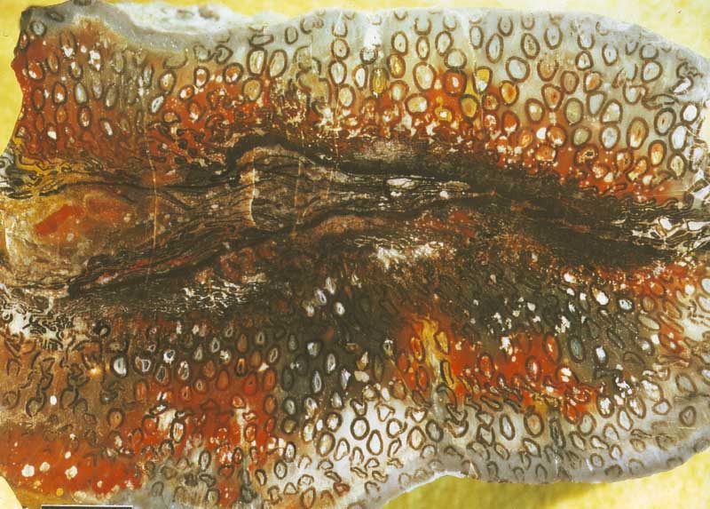
The specimen seen here is distinguished by an uncommon
combination of
advanced decay and excellent preservation: The collaps or squeezing
process which had affected virtually all
Psaronius
stems in the Döhlen Basin while lying
prostrate
in the swamp did not at all affect a large part of the aerial root
mantle of this stem. Only the centre and about
half the aerial root mantle became squeezed. Apparently the
tissues of the centre and adjacent roots became weakened by rot faster
than
influx of silica by diffusion could stabilize them so that the
centre and the roots near the centre collapsed.
Fig.1: Psaronius,
the stem of Scolecopteris,
the "maggot fern", in cross-section: most
beautiful specimen found hitherto in the Döhlen Basin. Width of the
picture 9cm.
Preservation by silicification
is governed by the
competition
between tissue decay and
silica uptake processes. These processes are
interdependent, which complicates things:
Silica influx is
precluded by diffusion barriers in healthy tissue and hence furthered
by progressive decay. On the other hand, decay becomes increasingly
delayed as silica accumulates in the tissue. Also the various tissues
are differentially resistant to decay. Considering all this, one need
not wonder why some features of the
fossil are not easily understood.
Apparently the silica penetrated the strong walls of the aerial roots
so slowly that
the inner tisse decayed and vanished before it could become
preserved by the mineral deposition.
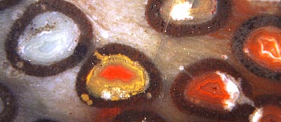
Silica subsequently entering into the cavity formed agates. Same as
with the agates in small cavities in volcanic rock, it is always a
cause of wonder that neighbouring ones may look completely different
from each other, as in Fig.2.
Obviously, tiny differences of the chemical constitution of the
water-filled cavities result in large effects on the
deposition of SiO2 and
colourful minerals, mostly iron oxides.
 Fig.2, left: Agates in hollow aerial roots.
  Width
of the picture 1.2cm.
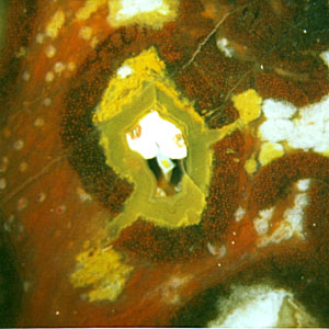
Fig.3,
right: Complex agate formed by a sequence of deposition processes in a
hollow aerial root after it had been split by a crack. Width of the
picture 0.5cm.
As a peculiar fact, the xylem strand of the aerial roots with its
characteristic "star-shaped" cross-section, which is usually preserved,
has vanished here together with the soft tissue surrounding it.
Contrary to
such complete decay of the interior of the aerial roots, the
cellular structure of their wall is completely
preserved. (Not well seen with this magnification.)
Sample: Bu7/24.2 , found at the type locality of Scolecopteris,
2000; Photographs: M. Barthel.
H.-J.
Weiss
2011
[1] R. Rößler :
Der versteinerte Wald von Chemnitz,
Naturkunde-Museum
Chemnitz, 2001.
|
 |
 6 6 |

 6
6
 Fig.2, left: Agates in hollow aerial roots.
  Width
of the picture 1.2cm.
 6
6