Various diffusion effects in
fossiliferous Permian chert
The omnipresent phenomenon of diffusion, whose essential part in
fossilisation is likely to be ignored by those who speak of circulating
solutions, has been vividly explained in a separate text. This
contribution is to draw the attention to a few of the various
manifestations of diffusion in chert.
Cracks
can influence diffusion
in contrary ways: They provide easy diffusion paths along but obstruct
diffusion across. Fig.1 shows a conspicuous case of a crack with an
opening so narrow that it is seen on the image as a thin line only but
nevertheless with a wide area of influence.
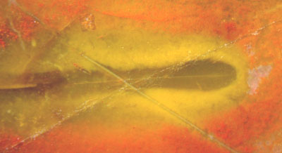
Fig.1:
Crack seen as a thin line with a wide halo resulting from diffusion,
incidentally crossed by two unrelated cracks without halo. Width of the
picture 4mm.
Apparently some agent coming along the
crack, probably by surface diffusion along the crack faces, caused
the small amout of hematite, which lends the red stain to the
chalzedony, to turn into the yellow goethite. Also it seems to have
reduced
part of the goethite to some colourless soluble iron compound which
probably escaped by diffusion.
The
configuration of the three nearly straight cracks in Fig.1 is
remarkable from a mechanical point of view: While propagating, they did
not feel each other's presence, hence they must have propagated at much
different times. The crack running from above left to below right is
the oldest one. Its 20µm opening is completely filled with silica so
that it does not act as a mechanical discontinuity. It must have been
so when the
horizontal crack came from the left and crossed it without the
slightest deflection. The horizontal
crack, too, must
have been healed up when the third crack crossed it without getting
deflected. (The third crack is still open, by which it has
been
recognized here as the third and last one.) Obviously, the
diffusion must have been going on while the second crack was open,
which is much later than the instant of the first cracking and a long
time before the instant of the third cracking. A very slight
deformation of the yellow and discoloured parts of the halo indicate
that the first crack slightly influenced the diffusion process although
it did not influence the path of the second crack.
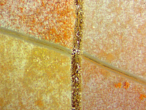
Fig.2: Cracks serving as diffusion barriers in chert with
granular precipitates
From
the even distribution of the precipitates of unknown nature on a small
part of the sample (Fig.2) it can be concluded that precipitation
came first. Next came the slanting crack, then healed up before the
perpendicular crack ran across bcause the latter did not feel the
former's presence.
It is not easy to reconstruct the sequence of
staining and /or bleaching of the precipitates. At least part of it
must have been going on when the second crack was there and already
filled with crystalline quartz as it is now. This follows from the
observation that the second crack did not serve as an easy
diffusion path. Hence one can assume, as the least complicated version,
that the diffusion barriers responsible for the different colours
did not consist in the cracks themselves but in some barrier function
of the solid fill of the healed-up cracks.
Apparently one cannot find out here whether it was staining or
bleaching which came with diffusion of some agent through the
chalzedony and was halted at the filled-up cracks. Again it appears
that there can be particular faces of discontinuity
in the chalzedony, as the faces of filled-up cracks
seen as lines on the cross-sections in Figs.1,2, which do not make
themselves felt mechanically but act as diffusion barriers only. The
details seem to be hidden in the molecular structure.
Other
small areas of the same sample section are suitable for contemplating
diffusion effects without interference by cracks. Fig.3 shows the same
type of precipitates as in Fig.2. The higher magnification apparently
does not convincingly suggest an explanation.
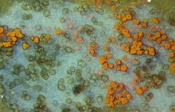
Fig.3: Enigmatic lumps of about 40µm in a milky patch of chalzedony.
Width of the picture 1.2mm.
Judging
from the evidence of bleaching in Fig.1 and possibly in Fig.2, the pale
lumps in Fig.3 could well have been bleached from red to transparent. A
conspicuous white spot in the middle of some bleached lumps might give
a clue. Unfortunately, more than one possible ways could have led to
what is seen here:
(1) A dissolving mineral grain in silica gel could have caused the
formation of the little lump
which became stained with hematite later.
(2) For reasons unknown, cyanobacteria could have grown preferably
around the white spot, thereby producing organic gel which
made the lump and oxygen which caused the precipitation
of hematite on the lump.
The latter interpretation is favoured
by the dark coating seen on some lumps, especially on the red ones. It
is possibly the latest layer of cyanobacteria,
which overgrew the
precipitate to get into the light, as it is known from cyanobacteria in
the Banded Iron Ore [1].
The pictures have been taken from one
chert sample found at the well-known site of fossiliferous pebbles and
boulders at Kleinnaundorf (Kohlenstr.), Freital, Döhlen Basin, Lower
Permian. This sample contains squeezed frond stalks and pinnules with
forked venation
and 4-fold synangia of the tree fern Scolecopteris,
also
moult parts of the crustacean Uronectes
and fungus
hyphae. Finally it is mentioned here that Scolecopteris,
the "maggot fern", is found abundantly in these cherts, debris from Uronectes moults
is less common, and fungus parts are rare.
Sample: found by H. Ahlheim, polished and provided by H. Albrecht,
Dresden, kept in the own collection under the label Bu7/207 .
H.-J. Weiss
2015
[1] T.N. Taylor
et al.: Paleobotany. Elsevier 2009
|
 |
 13 13 |