Permian tree fern details
Tree fern
foliage and stems make a major part of the fossils in the Permian
cherts found in the Doehlen basin, Germany. As a lucky coincidence,
pinnules of the big fronds appear on a small area of a cut face of a
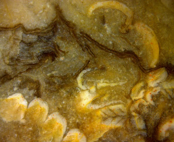
sample with various orientation: Fig.1.
Above right there is a pinna
cross-section with two pinnules cut lengthwise. The tilted cut of
the four 0.13mm-wide
pinnules below left makes them appear thicker than they really are.
Another pinnule is
cut such that its veins are clearly seen. Their angle of
about 45° with respect to
the midrib immediately leads to the dispute [1]
concerning the tree fern species in the Doehlen basin.
Fig.1: "Maggot fern" pinnules with various orientation. Image width
10mm.
As discussed
earlier [1], the repeatedly questioned
claim that there is only one tree fern species in the cherts
of the Doehlen basin [2], Scolecopteris elegans
known as "maggot fern", cannot be upheld.
Lately it has appeared that some of the tree
fern pinnules in chert offered as Sc. elegans including
even one alleged type specimen [3],
represent one ore more other species [4]. This newly arising
uncertainty concerning the "maggot fern" is one of the
reasons why this new contribution on an old
species seemingly well known since the early
days of paleobotany may be justified.
Fig.2 (below): "Maggot
fern" pinna cross-sections with pinnules cut lengthwise.
Note the sharp contour of the midrib of the above pinna. Image
width 10.5mm.
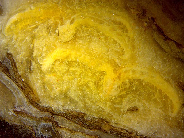
The confusion still prevailing in the scientific literature concerning
the "maggot fern" Scolecopteris elegans,
also known as Pecopteris
arborescens [5] or Scolecopteris arborescens [5] is
due to various reasons. One of them is the fact that the pinnule veins
are seldom seen as clearly as in Fig.1 and in [1]. Even among the
thousand pinnules on the magnificient image of the biggest "maggot
stone" ever found there is not one with clearly seen veins [2].
Aligned epidermis
cells had been mistaken for a dense cover of hairs on
the upper side of the pinnules ([5], p.280 and Tafel VIII Bild 7) but
this had been retracted later by the
author himself.
Scattered hairs may be present on the lower side of
the pinnule, as seen below right in Fig.2.
They
are easily confused with fungus hyphae but are recognized as hairs by
their attachment to the pinnule. For a clearer distinction of hyphae
and hairs see [6].
In view of the largely decayed tissue of the pinnules, the uncommonly
distinct contour of the midrib cross-section of
the above pinna in Fig.2 is enigmatic.
Fused sporangia, known as synangia, are not seen on cut faces here.
As a lucky coincidence, a slightly inclined cross-section
of a pinna on the raw surface of this sample (Fig.3) provides
a sideways view of a pinnule with a row of fringes seen from outside
and synangia hidden behind. Also it provides a lengthwise section of
the left pinnule with synangia.
With suitable position and orientation of the cut
plane, the fringes on
the pinnule margins can be conspicuous
on pinnule cross-sections<.
(See, for example, the logo at the bottom of this
page.)
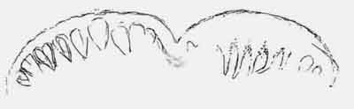
Fig.3: Slightly inclined cross-section of a pinna on the raw
surface of this sample, with fringes or lobes on the edge of the right
pinnule. Width of the image 8mm.
This small sample (0.3kg) with "maggot fern" is
distinguished among hundreds of others found in the Döhlen basin
not only by providing several details as seen in Figs.1-3 but also by
being the only one ever found near Pesterwitz. Repeated thorough search
on agricultural areas near Pesterwitz produced 14 mostly
small samples of
petrified wood, among them a unique one , not yet identified, and
a tree fern stem
fragment of the Psaronius
type (Fig.4). Since the "maggot
fern" stems are of the same type,
Fig.4 may serve as an illustratory addition to Figs.1-3.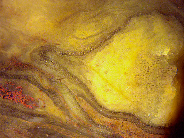
Fig.4: Psaronius
aerial roots with "star-shaped" cross-sections
of the central strand: with dainty cell walls (right),
or squeezed and
stained red with hematite (left), on the raw surface of a small sample of 41g. Image
width 7mm.
Psaronius
aerial roots preserved in chert are mostly seen in a more or less
squeezed state.
Hence, the well preserved central strand in this
sample, with clearly seen thin walls of big cells, is remarkable.
Samples found near
Pesterwitz, Doehlen basin; Figs.1-3: Pe/3 (1994); Fig.4: Pe/10 (1995).
H.-J. Weiss
2020
[1] Permian
Chert News 2 , Venation
pattern ...
[2] M.
Barthel: Die
Madensteine vom Windberg, Deutschland.
in: U.
Dernbach, W.D.
Tidwell:
Geheimnisse versteinerter Pflanzen. D'ORO Verlag, Heppenheim 2002,
p65-77.
[3] M.
Barthel: Die Rotliegend-Flora der Döhlen-Formation.
Geologica Saxonica 61(2) (2015), 108-229.
[4] Permian
Chert News 26 , Maggot
fern confusion.
[5] M.
Barthel: Pecopteris-Arten
v.
Schloiheims aus Typuslokalitäten
in der DDR.
Schriftenr. geolog. Wiss. - Berlin 16 (1980), 275-304.
[6] Permian
Chert News 17 ,
Microbes, fungi, maggot fern.
|
 |
 30 30 |