Persistent "palisade wall" of
Aglaophyton
sporangia
Sporangia of the early land plant Aglaophyton
are among those objects in the Lower Devonian Rhynie chert which are
most easily identified. While the identification of plant sections seen
on sample surfaces or cut chert faces is usually more difficult owing
to
shrivelling, squeezing, and rotting before fossilisation, Aglaophyton sporangia
are much less affected by such phenomena. Also they are distinguished
from any other plant parts (with exception of the rare Rhynia sporangia)
by an epidermis consisting of stacked slab-like
cells with rot-resistant walls which often appear as a row of palisades
on
capsule cross-sections, as seen here. The visual impression of separate
posts is also
due to the fact that the recurved parts of the cell walls, on
the surface of the capsule and inside, are most often not preserved,
for reasons unknown. (A rare exception is seen in Rhynie
Chert News 105.)
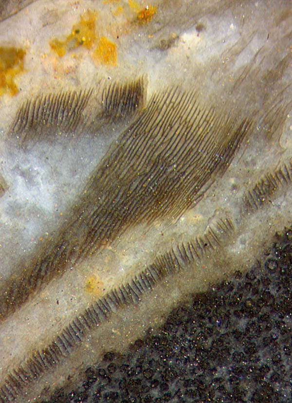
Figure: Palisade wall
fragments of Aglaophyton sporangium
in chert, rare
combination of parts with different aspect, degraded spores inside the
capsule below right. Image width 2mm.
Since incidental tangential cuts of the capsules are much less probable
than others, the capsule wall is seldom seen from outside. Hence the
fragments in this image offer a rare combination of different aspects
of the palisade wall.
Not seen here are two obvious deviations from the symmetry of the outer
shape of the capsule:
In Rhynie
Chert News 62
the narrow
cells are seen to be aligned with
a texture winding around the
spindle-shaped sporangium in a screw-like way.
Another unexpected asymmetry is hidden below the
surface but seen on cross-sections of the spore capsule: In
Rhynie
Chert News 65
the
orientation of the cell flanges appearing as
palisades is not always
radial as one should expect but varies along the circumference in some
systematic but not yet understood way.
No explanation can be given here for the combination of the axial
symmetry
of the spindle-shaped sporangia and the asymmetries of the epidermis
tissue
on their surface.
Sample: Rh9/69, 43g, Part 1, found by S.Weiss in 2004.
H.-J.
Weiss 2018
 |
 |
132 |